2XVZ

Cobalt chelatase CbiK (periplasmatic) from Desulvobrio vulgaris Hildenborough (co-crystallized with cobalt)


Experimental Data Snapshot

  • Method: X-RAY DIFFRACTION
  • Resolution: 2.40 Å
  • R-Value Free: 0.220 
  • R-Value Work: 0.170 
  • R-Value Observed: 0.172 

wwPDB Validation   3D Report Full Report


Ligand Structure Quality Assessment 


This is version 1.2 of the entry. See complete history


Literature

Evolution in a Family of Chelatases Facilitated by the Introduction of Active Site Asymmetry and Protein Oligomerization.

Romao, C.V.Ladakis, D.Lobo, S.A.L.Carrondo, M.A.Brindley, A.A.Deery, E.Matias, P.M.Pickersgill, R.W.Saraiva, L.M.Warren, M.J.

(2011) Proc Natl Acad Sci U S A 108: 97

  • DOI: https://doi.org/10.1073/pnas.1014298108
  • Primary Citation of Related Structures:  
    2XVX, 2XVZ, 2XWP, 2XWQ, 2XWS

  • PubMed Abstract: 

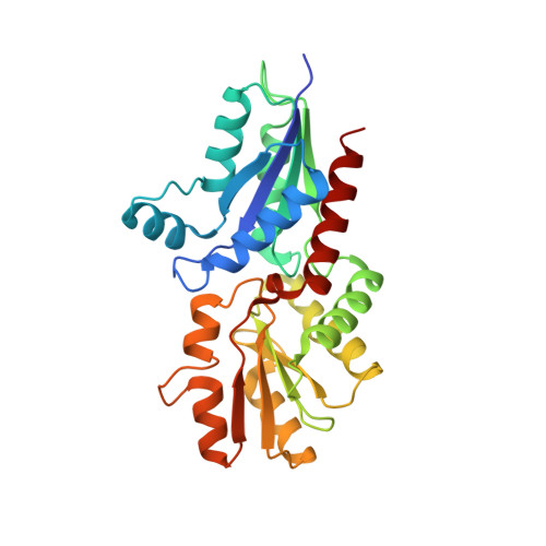
    The class II chelatases associated with heme, siroheme, and cobalamin biosynthesis are structurally related enzymes that insert a specific metal ion (Fe(2+) or Co(2+)) into the center of a modified tetrapyrrole (protoporphyrin or sirohydrochlorin). The structures of two related class II enzymes, CbiX(S) from Archaeoglobus fulgidus and CbiK from Salmonella enterica, that are responsible for the insertion of cobalt along the cobalamin biosynthesis pathway are presented in complex with their metallated product. A further structure of a CbiK from Desulfovibrio vulgaris Hildenborough reveals how cobalt is bound at the active site. The crystal structures show that the binding of sirohydrochlorin is distinctly different to porphyrin binding in the protoporphyrin ferrochelatases and provide a molecular overview of the mechanism of chelation. The structures also give insights into the evolution of chelatase form and function. Finally, the structure of a periplasmic form of Desulfovibrio vulgaris Hildenborough CbiK reveals a novel tetrameric arrangement of its subunits that are stabilized by the presence of a heme b cofactor. Whereas retaining colbaltochelatase activity, this protein has acquired a central cavity with the potential to chaperone or transport metals across the periplasmic space, thereby evolving a new use for an ancient protein subunit.


  • Organizational Affiliation

    Instituto de Tecnologia Química e Biológica, Universidade Nova de Lisboa, Avenida da República (EAN), 2780-157 Oeiras, Portugal. cmromao@itqb.unl.pt


Macromolecules
Find similar proteins by:  (by identity cutoff)  |  3D Structure
Entity ID: 1
MoleculeChains Sequence LengthOrganismDetailsImage
CHELATASE, PUTATIVE269Nitratidesulfovibrio vulgaris str. HildenboroughMutation(s): 0 
EC: 4.99.1.3
UniProt
Find proteins for Q72EC8 (Desulfovibrio vulgaris (strain ATCC 29579 / DSM 644 / NCIMB 8303 / VKM B-1760 / Hildenborough))
Explore Q72EC8 
Go to UniProtKB:  Q72EC8
Entity Groups  
Sequence Clusters30% Identity50% Identity70% Identity90% Identity95% Identity100% Identity
UniProt GroupQ72EC8
Sequence Annotations
Expand
  • Reference Sequence
Small Molecules
Ligands 7 Unique
IDChains Name / Formula / InChI Key2D Diagram3D Interactions
HEM
Query on HEM

Download Ideal Coordinates CCD File 
B [auth A]PROTOPORPHYRIN IX CONTAINING FE
C34 H32 Fe N4 O4
KABFMIBPWCXCRK-RGGAHWMASA-L
SO4
Query on SO4

Download Ideal Coordinates CCD File 
D [auth A]
E [auth A]
F [auth A]
J [auth A]
K [auth A]
D [auth A],
E [auth A],
F [auth A],
J [auth A],
K [auth A],
L [auth A],
M [auth A],
N [auth A]
SULFATE ION
O4 S
QAOWNCQODCNURD-UHFFFAOYSA-L
GOL
Query on GOL

Download Ideal Coordinates CCD File 
G [auth A],
H [auth A],
I [auth A]
GLYCEROL
C3 H8 O3
PEDCQBHIVMGVHV-UHFFFAOYSA-N
CO
Query on CO

Download Ideal Coordinates CCD File 
C [auth A]COBALT (II) ION
Co
XLJKHNWPARRRJB-UHFFFAOYSA-N
CL
Query on CL

Download Ideal Coordinates CCD File 
P [auth A]CHLORIDE ION
Cl
VEXZGXHMUGYJMC-UHFFFAOYSA-M
PER
Query on PER

Download Ideal Coordinates CCD File 
Q [auth A]PEROXIDE ION
O2
ANAIPYUSIMHBEL-UHFFFAOYSA-N
NA
Query on NA

Download Ideal Coordinates CCD File 
O [auth A]SODIUM ION
Na
FKNQFGJONOIPTF-UHFFFAOYSA-N
Experimental Data & Validation

Experimental Data

  • Method: X-RAY DIFFRACTION
  • Resolution: 2.40 Å
  • R-Value Free: 0.220 
  • R-Value Work: 0.170 
  • R-Value Observed: 0.172 
  • Space Group: I 4 2 2
Unit Cell:
Length ( Å )Angle ( ˚ )
a = 121.441α = 90
b = 121.441β = 90
c = 121.1γ = 90
Software Package:
Software NamePurpose
REFMACrefinement
XDSdata reduction
SCALEPACKdata scaling
SHELXphasing

Structure Validation

View Full Validation Report


Ligand Structure Quality Assessment 


Entry History 

Deposition Data

Revision History  (Full details and data files)

  • Version 1.0: 2010-12-22
    Type: Initial release
  • Version 1.1: 2011-05-08
    Changes: Version format compliance
  • Version 1.2: 2011-07-13
    Changes: Version format compliance