2XKA

Crystal structure of a GTPyS-form protofilament of Bacillus thuringiensis serovar israelensis TubZ


Experimental Data Snapshot

  • Method: X-RAY DIFFRACTION
  • Resolution: 3.00 Å
  • R-Value Free: 0.284 
  • R-Value Work: 0.232 
  • R-Value Observed: 0.235 

wwPDB Validation   3D Report Full Report


Ligand Structure Quality Assessment 


This is version 1.3 of the entry. See complete history


Literature

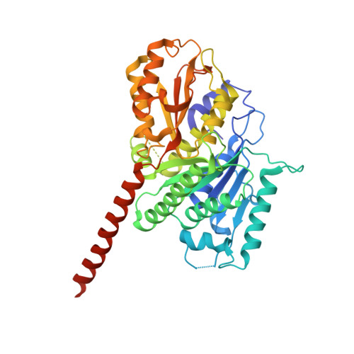
Filament Structure of Bacterial Tubulin Homologue Tubz.

Aylett, C.H.Wang, Q.Michie, K.A.Amos, L.A.Lowe, J.

(2010) Proc Natl Acad Sci U S A 107: 19766

  • DOI: https://doi.org/10.1073/pnas.1010176107
  • Primary Citation of Related Structures:  
    2XKA, 2XKB

  • PubMed Abstract: 

    Low copy number plasmids often depend on accurate partitioning systems for their continued survival. Generally, such systems consist of a centromere-like region of DNA, a DNA-binding adaptor, and a polymerizing cytomotive filament. Together these components drive newly replicated plasmids to opposite ends of the dividing cell. The Bacillus thuringiensis plasmid pBToxis relies on a filament of the tubulin/FtsZ-like protein TubZ for its segregation. By combining crystallography and electron microscopy, we have determined the structure of this filament. We explain how GTP hydrolysis weakens the subunit-subunit contact and also shed light on the partitioning of the plasmid-adaptor complex. The double helical superstructure of TubZ filaments is unusual for tubulin-like proteins. Filaments of ParM, the actin-like partitioning protein, are also double helical. We suggest that convergent evolution shapes these different types of cytomotive filaments toward a general mechanism for plasmid separation.


  • Organizational Affiliation

    Medical Research Council Laboratory of Molecular Biology, Cambridge CB2 0QH, United Kingdom.


Macromolecules
Find similar proteins by:  (by identity cutoff)  |  3D Structure
Entity ID: 1
MoleculeChains Sequence LengthOrganismDetailsImage
FTSZ/TUBULIN-RELATED PROTEIN
A, B, C, D, E
A, B, C, D, E, F, G
421Bacillus thuringiensis serovar israelensisMutation(s): 1 
UniProt
Find proteins for Q8KNP3 (Bacillus thuringiensis subsp. israelensis)
Explore Q8KNP3 
Go to UniProtKB:  Q8KNP3
Entity Groups  
Sequence Clusters30% Identity50% Identity70% Identity90% Identity95% Identity100% Identity
UniProt GroupQ8KNP3
Sequence Annotations
Expand
  • Reference Sequence
Small Molecules
Ligands 2 Unique
IDChains Name / Formula / InChI Key2D Diagram3D Interactions
GSP
Query on GSP

Download Ideal Coordinates CCD File 
H [auth A]
J [auth B]
L [auth C]
N [auth D]
P [auth E]
H [auth A],
J [auth B],
L [auth C],
N [auth D],
P [auth E],
R [auth F],
T [auth G]
5'-GUANOSINE-DIPHOSPHATE-MONOTHIOPHOSPHATE
C10 H16 N5 O13 P3 S
XOFLBQFBSOEHOG-UUOKFMHZSA-N
MG
Query on MG

Download Ideal Coordinates CCD File 
I [auth A]
K [auth B]
M [auth C]
O [auth D]
Q [auth E]
I [auth A],
K [auth B],
M [auth C],
O [auth D],
Q [auth E],
S [auth F],
U [auth G]
MAGNESIUM ION
Mg
JLVVSXFLKOJNIY-UHFFFAOYSA-N
Experimental Data & Validation

Experimental Data

  • Method: X-RAY DIFFRACTION
  • Resolution: 3.00 Å
  • R-Value Free: 0.284 
  • R-Value Work: 0.232 
  • R-Value Observed: 0.235 
  • Space Group: P 21 21 21
Unit Cell:
Length ( Å )Angle ( ˚ )
a = 51.06α = 90
b = 85.15β = 90
c = 632.23γ = 90
Software Package:
Software NamePurpose
MOSFLMdata reduction
SCALAdata scaling
SHELXphasing
SOLVEphasing
REFMACrefinement

Structure Validation

View Full Validation Report


Ligand Structure Quality Assessment 


Entry History 

Deposition Data

Revision History  (Full details and data files)

  • Version 1.0: 2010-11-10
    Type: Initial release
  • Version 1.1: 2011-05-08
    Changes: Version format compliance
  • Version 1.2: 2011-07-13
    Changes: Version format compliance
  • Version 1.3: 2018-01-24
    Changes: Source and taxonomy