2XB3

The Structure of Cyanobacterial PsbP


Experimental Data Snapshot

  • Method: X-RAY DIFFRACTION
  • Resolution: 2.80 Å
  • R-Value Free: 0.238 
  • R-Value Work: 0.192 
  • R-Value Observed: 0.194 

wwPDB Validation   3D Report Full Report


This is version 1.2 of the entry. See complete history


Literature

The Structure of Cyanop at 2.8A: Implications for the Evolution and Function of the Psbp Subunit of Photosystem II.

Michoux, F.Takasaka, K.Boehm, M.Nixon, P.Murray, J.W.

(2010) Biochemistry 49: 7411

  • DOI: https://doi.org/10.1021/bi1011145
  • Primary Citation of Related Structures:  
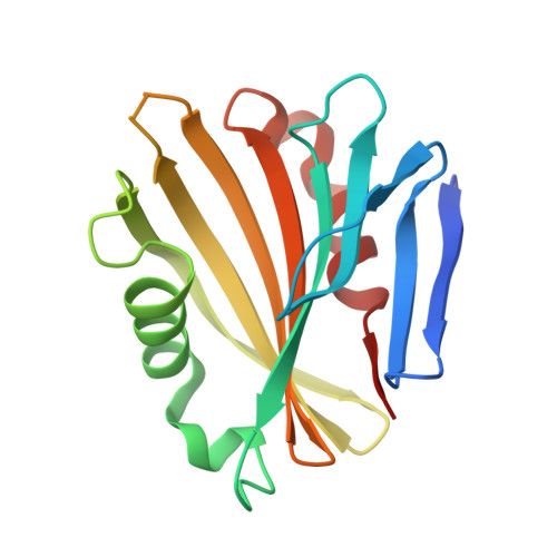
    2XB3

  • PubMed Abstract: 

    We present here the crystal structure of CyanoP (Tlr2075) from Thermosynechococcus elongatus at 2.8 A. CyanoP is a substoichiometric component of the isolated cyanobacterial Photosystem II (PSII) complex, distantly related to the PsbP extrinsic subunit of the oxygen-evolving PSII complex in higher plants and green algae. Despite the relatively low degree of sequence similarity, we have found that CyanoP adopts the same beta-sandwich fold as higher-plant PsbP and contains a well-conserved metal (zinc)-binding site that is also present in plant PsbP. Our results support the idea that CyanoP represents the basal structural fold of the PsbP superfamily.


  • Organizational Affiliation

    Division of Biology, Wolfson Biochemistry Building, Imperial College London,South Kensington Campus, London SW7 2AZ, UK.


Macromolecules
Find similar proteins by:  (by identity cutoff)  |  3D Structure
Entity ID: 1
MoleculeChains Sequence LengthOrganismDetailsImage
PSBP PROTEIN165Thermosynechococcus vestitus BP-1Mutation(s): 0 
UniProt
Find proteins for Q8DH84 (Thermosynechococcus vestitus (strain NIES-2133 / IAM M-273 / BP-1))
Explore Q8DH84 
Go to UniProtKB:  Q8DH84
Entity Groups  
Sequence Clusters30% Identity50% Identity70% Identity90% Identity95% Identity100% Identity
UniProt GroupQ8DH84
Sequence Annotations
Expand
  • Reference Sequence
Experimental Data & Validation

Experimental Data

  • Method: X-RAY DIFFRACTION
  • Resolution: 2.80 Å
  • R-Value Free: 0.238 
  • R-Value Work: 0.192 
  • R-Value Observed: 0.194 
  • Space Group: P 32 2 1
Unit Cell:
Length ( Å )Angle ( ˚ )
a = 64.946α = 90
b = 64.946β = 90
c = 81.806γ = 120
Software Package:
Software NamePurpose
REFMACrefinement
MOSFLMdata reduction
SCALAdata scaling
PHENIXphasing

Structure Validation

View Full Validation Report


Entry History 

Deposition Data

Revision History  (Full details and data files)

  • Version 1.0: 2010-08-25
    Type: Initial release
  • Version 1.1: 2011-05-08
    Changes: Version format compliance
  • Version 1.2: 2011-07-13
    Changes: Version format compliance