2X3H

COLIPHAGE K5A LYASE


Experimental Data Snapshot

  • Method: X-RAY DIFFRACTION
  • Resolution: 1.60 Å
  • R-Value Free: 0.203 
  • R-Value Work: 0.176 
  • R-Value Observed: 0.178 

wwPDB Validation   3D Report Full Report


This is version 1.4 of the entry. See complete history


Literature

The K5 Lyase Kfla Combines a Viral Tail Spike Structure with a Bacterial Polysaccharide Lyase Mechanism.

Thompson, J.E.Pourhossein, M.Waterhouse, A.Hudson, T.Goldrick, M.Derrick, J.P.Roberts, I.S.

(2010) J Biol Chem 285: 23963

  • DOI: https://doi.org/10.1074/jbc.M110.127571
  • Primary Citation of Related Structures:  
    2X3H

  • PubMed Abstract: 

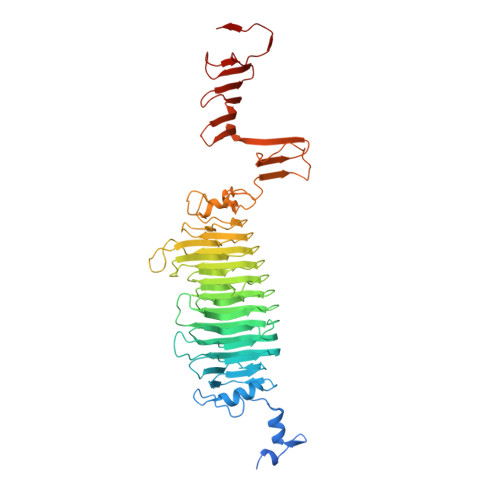
    K5 lyase A (KflA) is a tail spike protein (TSP) encoded by a K5A coliphage, which cleaves K5 capsular polysaccharide, a glycosaminoglycan with the repeat unit [-4)-betaGlcA-(1,4)- alphaGlcNAc(1-], displayed on the surface of Escherichia coli K5 strains. The crystal structure of KflA reveals a trimeric arrangement, with each monomer containing a right-handed, single-stranded parallel beta-helix domain. Stable trimer formation by the intertwining of strands in the C-terminal domain, followed by proteolytic maturation, is likely to be catalyzed by an autochaperone as described for K1F endosialidase. The structure of KflA represents the first bacteriophage tail spike protein combining polysaccharide lyase activity with a single-stranded parallel beta-helix fold. We propose a catalytic site and mechanism representing convergence with the syn-beta-elimination site of heparinase II from Pedobacter heparinus.


  • Organizational Affiliation

    Faculty of Life Sciences, University of Manchester, Manchester M13 9PT, United Kingdom.


Macromolecules
Find similar proteins by:  (by identity cutoff)  |  3D Structure
Entity ID: 1
MoleculeChains Sequence LengthOrganismDetailsImage
K5 LYASE
A, B, C
542Vectrevirus K15Mutation(s): 0 
EC: 4.2.2.7
UniProt
Find proteins for O09496 (Escherichia virus K5)
Explore O09496 
Go to UniProtKB:  O09496
Entity Groups  
Sequence Clusters30% Identity50% Identity70% Identity90% Identity95% Identity100% Identity
UniProt GroupO09496
Sequence Annotations
Expand
  • Reference Sequence
Small Molecules
Ligands 1 Unique
IDChains Name / Formula / InChI Key2D Diagram3D Interactions
BR
Query on BR

Download Ideal Coordinates CCD File 
D [auth A]
E [auth A]
F [auth A]
G [auth A]
H [auth A]
D [auth A],
E [auth A],
F [auth A],
G [auth A],
H [auth A],
I [auth B],
J [auth B],
K [auth C],
L [auth C],
M [auth C],
N [auth C],
O [auth C]
BROMIDE ION
Br
CPELXLSAUQHCOX-UHFFFAOYSA-M
Experimental Data & Validation

Experimental Data

  • Method: X-RAY DIFFRACTION
  • Resolution: 1.60 Å
  • R-Value Free: 0.203 
  • R-Value Work: 0.176 
  • R-Value Observed: 0.178 
  • Space Group: P 21 21 21
Unit Cell:
Length ( Å )Angle ( ˚ )
a = 68.53α = 90
b = 130.4β = 90
c = 145.74γ = 90
Software Package:
Software NamePurpose
REFMACrefinement
MOSFLMdata reduction
SCALAdata scaling
SOLVEphasing

Structure Validation

View Full Validation Report


Entry History 

Deposition Data

Revision History  (Full details and data files)

  • Version 1.0: 2010-06-02
    Type: Initial release
  • Version 1.1: 2011-05-07
    Changes: Version format compliance
  • Version 1.2: 2011-07-13
    Changes: Version format compliance
  • Version 1.3: 2019-05-08
    Changes: Data collection, Derived calculations, Experimental preparation, Other
  • Version 1.4: 2019-10-30
    Changes: Advisory, Data collection, Derived calculations, Other