2WKW

Alcaligenes esterase complexed with product analogue


Experimental Data Snapshot

  • Method: X-RAY DIFFRACTION
  • Resolution: 2.03 Å
  • R-Value Free: 0.194 
  • R-Value Work: 0.155 
  • R-Value Observed: 0.157 

wwPDB Validation   3D Report Full Report


Ligand Structure Quality Assessment 


This is version 1.3 of the entry. See complete history


Literature

The Atomic-Resolution Structure of a Novel Bacterial Esterase.

Bourne, P.C.Isupov, M.N.Littlechild, J.A.

(2000) Structure 8: 143

  • DOI: https://doi.org/10.1016/s0969-2126(00)00090-3
  • Primary Citation of Related Structures:  
    1QLW, 2WKW

  • PubMed Abstract: 

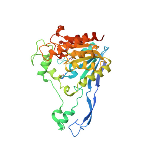
    A novel bacterial esterase that cleaves esters on halogenated cyclic compounds has been isolated from an Alcaligenes species. This esterase 713 is encoded by a 1062 base pair gene. The presence of a leader sequence of 27 amino acids suggests that this enzyme is exported from the cytosol. Esterase 713 has been over-expressed in Agrobacterium without this leader sequence. Its amino acid sequence shows no significant homology to any known protein sequence.


  • Organizational Affiliation

    Schools of Chemistry and Biological Sciences, University of Exeter, Exeter, EX4 4QD, UK.


Macromolecules
Find similar proteins by:  (by identity cutoff)  |  3D Structure
Entity ID: 1
MoleculeChains Sequence LengthOrganismDetailsImage
CARBOXYLESTERASE
A, B
328Alcaligenes sp.Mutation(s): 0 
UniProt
Find proteins for Q7SIA5 (Alcaligenes sp)
Explore Q7SIA5 
Go to UniProtKB:  Q7SIA5
Entity Groups  
Sequence Clusters30% Identity50% Identity70% Identity90% Identity95% Identity100% Identity
UniProt GroupQ7SIA5
Sequence Annotations
Expand
  • Reference Sequence
Small Molecules
Ligands 3 Unique
IDChains Name / Formula / InChI Key2D Diagram3D Interactions
W22
Query on W22

Download Ideal Coordinates CCD File 
C [auth A],
L [auth B]
[(2S)-4-methyl-3-oxo-2,3,4,5-tetrahydro-1H-1,4-benzodiazepin-2-yl]acetic acid
C12 H14 N2 O3
CLWDLBDPVUWYEW-JTQLQIEISA-N
SO4
Query on SO4

Download Ideal Coordinates CCD File 
D [auth A]
E [auth A]
I [auth A]
J [auth A]
M [auth B]
D [auth A],
E [auth A],
I [auth A],
J [auth A],
M [auth B],
P [auth B],
R [auth B]
SULFATE ION
O4 S
QAOWNCQODCNURD-UHFFFAOYSA-L
GOL
Query on GOL

Download Ideal Coordinates CCD File 
F [auth A]
G [auth A]
H [auth A]
K [auth A]
N [auth B]
F [auth A],
G [auth A],
H [auth A],
K [auth A],
N [auth B],
O [auth B],
Q [auth B]
GLYCEROL
C3 H8 O3
PEDCQBHIVMGVHV-UHFFFAOYSA-N
Experimental Data & Validation

Experimental Data

  • Method: X-RAY DIFFRACTION
  • Resolution: 2.03 Å
  • R-Value Free: 0.194 
  • R-Value Work: 0.155 
  • R-Value Observed: 0.157 
  • Space Group: P 21 21 21
Unit Cell:
Length ( Å )Angle ( ˚ )
a = 57.597α = 90
b = 115.811β = 90
c = 131.144γ = 90
Software Package:
Software NamePurpose
REFMACrefinement
DENZOdata reduction
SCALEPACKdata scaling
AMoREphasing

Structure Validation

View Full Validation Report


Ligand Structure Quality Assessment 


Entry History 

Deposition Data

Revision History  (Full details and data files)

  • Version 1.0: 2009-06-30
    Type: Initial release
  • Version 1.1: 2014-02-05
    Changes: Database references, Derived calculations, Non-polymer description, Other, Version format compliance
  • Version 1.2: 2019-07-24
    Changes: Data collection
  • Version 1.3: 2023-12-13
    Changes: Data collection, Database references, Derived calculations, Other, Refinement description