2WAG

The Structure of a family 25 Glycosyl hydrolase from Bacillus anthracis.


Experimental Data Snapshot

  • Method: X-RAY DIFFRACTION
  • Resolution: 1.40 Å
  • R-Value Free: 0.212 
  • R-Value Work: 0.187 
  • R-Value Observed: 0.189 

wwPDB Validation   3D Report Full Report


This is version 1.4 of the entry. See complete history


Literature

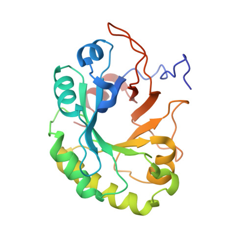
The Crystal Structure of a Family Gh25 Lysozyme from Bacillus Anthracis Implies a Neighboring-Group Catalytic Mechanism with Retention of Anomeric Configuration

Martinez-Fleites, C.Korczynska, J.E.Cope, M.Turkenburg, J.P.Taylor, E.J.

(2009) Carbohydr Res 344: 1753

  • DOI: https://doi.org/10.1016/j.carres.2009.06.001
  • Primary Citation of Related Structures:  
    2WAG

  • PubMed Abstract: 

    Lysozymes are found in many of the sequence-based families of glycoside hydrolases (www.cazy.org) where they show considerable structural and mechanistic diversity. Lysozymes from glycoside hydrolase family GH25 adopt a (alpha/beta)(5)(beta)(3)-barrel-like fold with a proposal in the literature that these enzymes act with inversion of anomeric configuration; the lack of a suitable substrate, however, means that no group has successfully demonstrated the configuration of the product. Here we report the 3-D structure of the GH25 enzyme from Bacillus anthracis at 1.4A resolution. We show that the active center is extremely similar to those from glycoside hydrolase families GH18, GH20, GH56, GH84, and GH85 implying that, in the absence of evidence to the contrary, GH25 enzymes also act with net retention of anomeric configuration using the neighboring-group catalytic mechanism that is common to this 'super-family' of enzymes.


  • Organizational Affiliation

    Structural Biology Laboratory, Department of Chemistry, The University of York, YO10 5YW, United Kingdom.


Macromolecules
Find similar proteins by:  (by identity cutoff)  |  3D Structure
Entity ID: 1
MoleculeChains Sequence LengthOrganismDetailsImage
LYSOZYME, PUTATIVE220Bacillus anthracis str. AmesMutation(s): 0 
EC: 3.2.1.17
UniProt
Find proteins for A0A6L7H2K3 (Bacillus anthracis)
Explore A0A6L7H2K3 
Go to UniProtKB:  A0A6L7H2K3
Entity Groups  
Sequence Clusters30% Identity50% Identity70% Identity90% Identity95% Identity100% Identity
UniProt GroupA0A6L7H2K3
Sequence Annotations
Expand
  • Reference Sequence
Small Molecules
Ligands 4 Unique
IDChains Name / Formula / InChI Key2D Diagram3D Interactions
15P
Query on 15P

Download Ideal Coordinates CCD File 
B [auth A],
J [auth A],
K [auth A],
L [auth A],
M [auth A]
POLYETHYLENE GLYCOL (N=34)
C69 H140 O35
VUYXVWGKCKTUMF-UHFFFAOYSA-N
SO4
Query on SO4

Download Ideal Coordinates CCD File 
C [auth A],
D [auth A],
E [auth A],
F [auth A],
G [auth A]
SULFATE ION
O4 S
QAOWNCQODCNURD-UHFFFAOYSA-L
GOL
Query on GOL

Download Ideal Coordinates CCD File 
H [auth A],
I [auth A]
GLYCEROL
C3 H8 O3
PEDCQBHIVMGVHV-UHFFFAOYSA-N
MG
Query on MG

Download Ideal Coordinates CCD File 
N [auth A]MAGNESIUM ION
Mg
JLVVSXFLKOJNIY-UHFFFAOYSA-N
Experimental Data & Validation

Experimental Data

  • Method: X-RAY DIFFRACTION
  • Resolution: 1.40 Å
  • R-Value Free: 0.212 
  • R-Value Work: 0.187 
  • R-Value Observed: 0.189 
  • Space Group: P 61 2 2
Unit Cell:
Length ( Å )Angle ( ˚ )
a = 100.522α = 90
b = 100.521β = 90
c = 106.72γ = 120
Software Package:
Software NamePurpose
REFMACrefinement
MOSFLMdata reduction
SCALAdata scaling
AMoREphasing

Structure Validation

View Full Validation Report


Entry History 

Deposition Data

Revision History  (Full details and data files)

  • Version 1.0: 2009-06-23
    Type: Initial release
  • Version 1.1: 2011-05-08
    Changes: Version format compliance
  • Version 1.2: 2011-07-13
    Changes: Version format compliance
  • Version 1.3: 2019-10-16
    Changes: Advisory, Data collection, Experimental preparation, Other
  • Version 1.4: 2023-12-13
    Changes: Advisory, Data collection, Database references, Derived calculations, Refinement description