


Discovery and Initial Sar of Quinazoline Inhibitors of Glmu from Haemophilus Influenzae
Melnick, M., Mochalkin, I., Lightle, S., Narasimhan, L., Mcdowell, L., Sarver, R.To be published.
Experimental Data Snapshot
Entity ID: 1 | |||||
|---|---|---|---|---|---|
| Molecule | Chains | Sequence Length | Organism | Details | Image |
| GLUCOSAMINE-1-PHOSPHATE N-ACETYLTRANSFERASE | 456 | Haemophilus influenzae | Mutation(s): 0 EC: 2.3.1.157 | 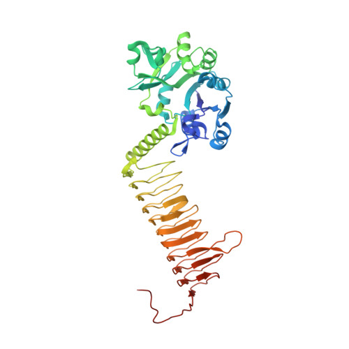 | |
UniProt | |||||
Find proteins for P43889 (Haemophilus influenzae (strain ATCC 51907 / DSM 11121 / KW20 / Rd)) Explore P43889 Go to UniProtKB: P43889 | |||||
Entity Groups | |||||
| Sequence Clusters | 30% Identity50% Identity70% Identity90% Identity95% Identity100% Identity | ||||
| UniProt Group | P43889 | ||||
Sequence AnnotationsExpand | |||||
| |||||
| Ligands 4 Unique | |||||
|---|---|---|---|---|---|
| ID | Chains | Name / Formula / InChI Key | 2D Diagram | 3D Interactions | |
| LZR Query on LZR | B [auth A] | 6-(CYCLOPROP-2-EN-1-YLMETHOXY)-2-[6-(CYCLOPROPYLMETHYL)-5-OXO-3,4,5,6-TETRAHYDRO-2,6-NAPHTHYRIDIN-2(1H)-YL]-7-METHOXYQUINAZOLIN-4(3H)-ONE C25 H26 N4 O4 BVRCGDWCYYETOO-UHFFFAOYSA-N |  | ||
| PG4 Query on PG4 | C [auth A] | TETRAETHYLENE GLYCOL C8 H18 O5 UWHCKJMYHZGTIT-UHFFFAOYSA-N |  | ||
| PGE Query on PGE | D [auth A], E [auth A] | TRIETHYLENE GLYCOL C6 H14 O4 ZIBGPFATKBEMQZ-UHFFFAOYSA-N |  | ||
| SO4 Query on SO4 | F [auth A] G [auth A] H [auth A] I [auth A] J [auth A] | SULFATE ION O4 S QAOWNCQODCNURD-UHFFFAOYSA-L |  | ||
| Length ( Å ) | Angle ( ˚ ) |
|---|---|
| a = 108.821 | α = 90 |
| b = 108.821 | β = 90 |
| c = 326.85 | γ = 120 |
| Software Name | Purpose |
|---|---|
| REFMAC | refinement |
| HKL-2000 | data reduction |
| HKL-2000 | data scaling |
| MOLREP | phasing |
RCSB PDB (citation) is hosted by
RCSB PDB is a member of the

