2W0U

CRYSTAL STRUCTURE OF HUMAN GLYCOLATE OXIDASE IN COMPLEX WITH THE INHIBITOR 5-[(4-CHLOROPHENYL)SULFANYL]- 1,2,3-THIADIAZOLE-4-CARBOXYLATE.


Experimental Data Snapshot

  • Method: X-RAY DIFFRACTION
  • Resolution: 2.84 Å
  • R-Value Free: 0.250 
  • R-Value Work: 0.205 
  • R-Value Observed: 0.207 

wwPDB Validation   3D Report Full Report


Ligand Structure Quality Assessment 


This is version 1.4 of the entry. See complete history


Literature

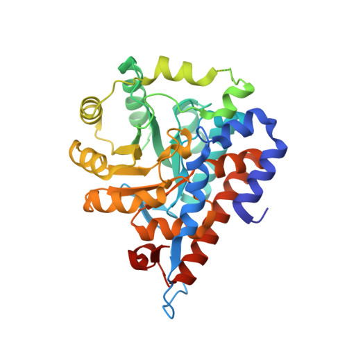
Structure of Human Glycolate Oxidase in Complex with the Inhibitor 4-Carboxy-5-[(4-Chlorophenyl)Sulfanyl]-1,2,3-Thiadiazole.

Bourhis, J.M.Vignaud, C.Pietrancosta, N.Gueritte, F.Guenard, D.Lederer, F.Lindqvist, Y.

(2009) Acta Crystallogr Sect F Struct Biol Cryst Commun 65: 1246

  • DOI: https://doi.org/10.1107/S1744309109041670
  • Primary Citation of Related Structures:  
    2W0U

  • PubMed Abstract: 

    Glycolate oxidase, a peroxisomal flavoenzyme, generates glyoxylate at the expense of oxygen. When the normal metabolism of glyoxylate is impaired by the mutations that are responsible for the genetic diseases hyperoxaluria types 1 and 2, glyoxylate yields oxalate, which forms insoluble calcium deposits, particularly in the kidneys. Glycolate oxidase could thus be an interesting therapeutic target. The crystal structure of human glycolate oxidase (hGOX) in complex with 4-carboxy-5-[(4-chlorophenyl)sulfanyl]-1,2,3-thiadiazole (CCPST) has been determined at 2.8 A resolution. The inhibitor heteroatoms interact with five active-site residues that have been implicated in catalysis in homologous flavodehydrogenases of L-2-hydroxy acids. In addition, the chlorophenyl substituent is surrounded by nonconserved hydrophobic residues. The present study highlights the role of mobility in ligand binding by glycolate oxidase. In addition, it pinpoints several structural differences between members of the highly conserved family of flavodehydrogenases of L-2-hydroxy acids.


  • Organizational Affiliation

    Department of Medical Biochemistry and Biophysics, Karolinska Institutet, S-171 77 Stockholm, Sweden.


Macromolecules
Find similar proteins by:  (by identity cutoff)  |  3D Structure
Entity ID: 1
MoleculeChains Sequence LengthOrganismDetailsImage
HYDROXYACID OXIDASE 1
A, B, C, D
370Homo sapiensMutation(s): 0 
EC: 1.1.3.15
UniProt & NIH Common Fund Data Resources
Find proteins for Q9UJM8 (Homo sapiens)
Explore Q9UJM8 
Go to UniProtKB:  Q9UJM8
PHAROS:  Q9UJM8
GTEx:  ENSG00000101323 
Entity Groups  
Sequence Clusters30% Identity50% Identity70% Identity90% Identity95% Identity100% Identity
UniProt GroupQ9UJM8
Sequence Annotations
Expand
  • Reference Sequence
Experimental Data & Validation

Experimental Data

  • Method: X-RAY DIFFRACTION
  • Resolution: 2.84 Å
  • R-Value Free: 0.250 
  • R-Value Work: 0.205 
  • R-Value Observed: 0.207 
  • Space Group: P 4 21 2
Unit Cell:
Length ( Å )Angle ( ˚ )
a = 138.69α = 90
b = 138.69β = 90
c = 186.84γ = 90
Software Package:
Software NamePurpose
REFMACrefinement
XDSdata reduction
SCALAdata scaling
MOLREPphasing

Structure Validation

View Full Validation Report


Ligand Structure Quality Assessment 


Entry History 

Deposition Data

Revision History  (Full details and data files)

  • Version 1.0: 2009-11-10
    Type: Initial release
  • Version 1.1: 2011-05-08
    Changes: Version format compliance
  • Version 1.2: 2011-07-13
    Changes: Version format compliance
  • Version 1.3: 2019-05-08
    Changes: Data collection, Experimental preparation, Other
  • Version 1.4: 2023-12-13
    Changes: Data collection, Database references, Derived calculations, Other, Refinement description