2W00

Crystal structure of the HsdR subunit of the EcoR124I restriction enzyme in complex with ATP


Experimental Data Snapshot

  • Method: X-RAY DIFFRACTION
  • Resolution: 2.60 Å
  • R-Value Free: 0.265 
  • R-Value Work: 0.221 
  • R-Value Observed: 0.221 

wwPDB Validation   3D Report Full Report


Ligand Structure Quality Assessment 


This is version 1.4 of the entry. See complete history

Re-refinement Note

A newer entry is available that reflects an alternative modeling of the original data: 6H2J


Literature

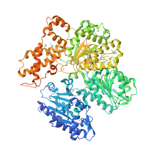
Structure of the Motor Subunit of Type I Restriction-Modification Complex Ecor124I.

Lapkouski, M.Panjikar, S.Janscak, P.Smatanova, I.K.Carey, J.Ettrich, R.Csefalvay, E.

(2009) Nat Struct Mol Biol 16: 94

  • DOI: https://doi.org/10.1038/nsmb.1523
  • Primary Citation of Related Structures:  
    2W00

  • PubMed Abstract: 

    Type I restriction-modification enzymes act as conventional adenine methylases on hemimethylated DNAs, but unmethylated recognition targets induce them to translocate thousands of base pairs before cleaving distant sites nonspecifically. The first crystal structure of a type I motor subunit responsible for translocation and cleavage suggests how the pentameric translocating complex is assembled and provides a structural framework for translocation of duplex DNA by RecA-like ATPase motors.


  • Organizational Affiliation

    Department of Structure and Function of Proteins, Institute of Systems Biology and Ecology, Academy of Sciences of the Czech Republic, Nove Hrady, Czech Republic.


Macromolecules
Find similar proteins by:  (by identity cutoff)  |  3D Structure
Entity ID: 1
MoleculeChains Sequence LengthOrganismDetailsImage
HSDR
A, B
1,038Escherichia coliMutation(s): 0 
EC: 3.1.21.3
UniProt
Find proteins for P10486 (Escherichia coli)
Explore P10486 
Go to UniProtKB:  P10486
Entity Groups  
Sequence Clusters30% Identity50% Identity70% Identity90% Identity95% Identity100% Identity
UniProt GroupP10486
Sequence Annotations
Expand
  • Reference Sequence
Small Molecules
Experimental Data & Validation

Experimental Data

  • Method: X-RAY DIFFRACTION
  • Resolution: 2.60 Å
  • R-Value Free: 0.265 
  • R-Value Work: 0.221 
  • R-Value Observed: 0.221 
  • Space Group: P 21 21 21
Unit Cell:
Length ( Å )Angle ( ˚ )
a = 123.91α = 90
b = 129.922β = 90
c = 161.01γ = 90
Software Package:
Software NamePurpose
REFMACrefinement
DENZOdata reduction
SCALEPACKdata scaling
Auto-Rickshawphasing

Structure Validation

View Full Validation Report


Ligand Structure Quality Assessment 


Entry History 

Deposition Data

Revision History  (Full details and data files)

  • Version 1.0: 2008-12-16
    Type: Initial release
  • Version 1.1: 2011-07-13
    Changes: Advisory, Version format compliance
  • Version 1.2: 2019-03-06
    Changes: Data collection, Derived calculations, Experimental preparation, Other
  • Version 1.3: 2019-05-08
    Changes: Data collection, Experimental preparation
  • Version 1.4: 2019-07-24
    Changes: Data collection