2VY0

The X-ray structure of endo-beta-1,3-glucanase from Pyrococcus furiosus


Experimental Data Snapshot

  • Method: X-RAY DIFFRACTION
  • Resolution: 2.16 Å
  • R-Value Free: 0.229 
  • R-Value Work: 0.191 
  • R-Value Observed: 0.193 

wwPDB Validation   3D Report Full Report


This is version 1.3 of the entry. See complete history


Literature

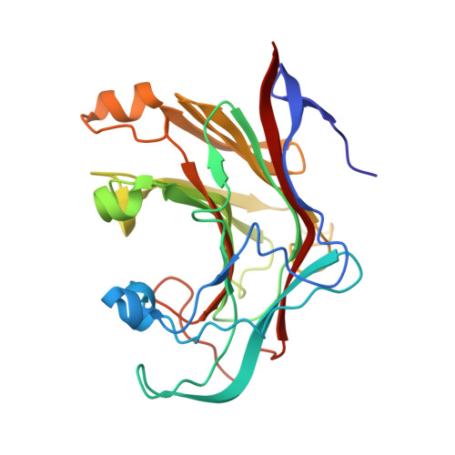
Crystal Structure of a Family 16 Endoglucanase from the Hyperthermophile Pyrococcus Furiosus-Structural Basis of Substrate Recognition.

Ilari, A.Fiorillo, A.Angelaccio, S.Florio, R.Chiaraluce, R.Van Der Oost, J.Consalvi, V.

(2009) FEBS J 276: 1048

  • DOI: https://doi.org/10.1111/j.1742-4658.2008.06848.x
  • Primary Citation of Related Structures:  
    2VY0

  • PubMed Abstract: 

    Bacterial and archaeal endo-beta-1,3-glucanases that belong to glycoside hydrolase family 16 share a beta-jelly-roll fold, but differ significantly in sequence and in substrate specificity. The crystal structure of the laminarinase (EC 3.2.1.39) from the hyperthermophilic archaeon Pyrococcus furiosus (pfLamA) has been determined at 2.1 A resolution by molecular replacement. The pfLamA structure reveals a kink of six residues (72-77) at the entrance of the catalytic cleft. This peptide is absent in the endoglucanases from alkaliphilic Nocardiopsis sp. strain F96 and Bacillus macerans, two proteins displaying an overall fold similar to that of pfLamA, but with different substrate specificity. A deletion mutant of pfLamA, lacking residues 72-75, hydrolyses the mixed-linkage beta-1,3-1,4-glucan lichenan 10 times more efficiently than the wild-type protein, indicating the importance of the kink in substrate preference.


  • Organizational Affiliation

    CNR Institute of Molecular Biology and Pathology, Italy. andrea.ilari@uniroma1.it


Macromolecules
Find similar proteins by:  (by identity cutoff)  |  3D Structure
Entity ID: 1
MoleculeChains Sequence LengthOrganismDetailsImage
ENDO-BETA-1,3-GLUCANASE
A, B
264Pyrococcus furiosusMutation(s): 0 
EC: 3.2.1.39
UniProt
Find proteins for O73951 (Pyrococcus furiosus)
Explore O73951 
Go to UniProtKB:  O73951
Entity Groups  
Sequence Clusters30% Identity50% Identity70% Identity90% Identity95% Identity100% Identity
UniProt GroupO73951
Sequence Annotations
Expand
  • Reference Sequence
Small Molecules
Ligands 5 Unique
IDChains Name / Formula / InChI Key2D Diagram3D Interactions
MRD
Query on MRD

Download Ideal Coordinates CCD File 
H [auth A],
N [auth B]
(4R)-2-METHYLPENTANE-2,4-DIOL
C6 H14 O2
SVTBMSDMJJWYQN-RXMQYKEDSA-N
MPD
Query on MPD

Download Ideal Coordinates CCD File 
F [auth A],
G [auth A]
(4S)-2-METHYL-2,4-PENTANEDIOL
C6 H14 O2
SVTBMSDMJJWYQN-YFKPBYRVSA-N
CA
Query on CA

Download Ideal Coordinates CCD File 
E [auth A],
J [auth B]
CALCIUM ION
Ca
BHPQYMZQTOCNFJ-UHFFFAOYSA-N
CL
Query on CL

Download Ideal Coordinates CCD File 
C [auth A],
I [auth B],
M [auth B]
CHLORIDE ION
Cl
VEXZGXHMUGYJMC-UHFFFAOYSA-M
NA
Query on NA

Download Ideal Coordinates CCD File 
D [auth A],
K [auth B],
L [auth B]
SODIUM ION
Na
FKNQFGJONOIPTF-UHFFFAOYSA-N
Experimental Data & Validation

Experimental Data

  • Method: X-RAY DIFFRACTION
  • Resolution: 2.16 Å
  • R-Value Free: 0.229 
  • R-Value Work: 0.191 
  • R-Value Observed: 0.193 
  • Space Group: P 1 21 1
Unit Cell:
Length ( Å )Angle ( ˚ )
a = 44.363α = 90
b = 84.762β = 104.97
c = 69.232γ = 90
Software Package:
Software NamePurpose
REFMACrefinement
DENZOdata reduction
SCALEPACKdata scaling
MOLREPphasing

Structure Validation

View Full Validation Report


Entry History 

Deposition Data

Revision History  (Full details and data files)

  • Version 1.0: 2009-03-24
    Type: Initial release
  • Version 1.1: 2011-05-07
    Changes: Version format compliance
  • Version 1.2: 2011-07-13
    Changes: Version format compliance
  • Version 1.3: 2023-12-13
    Changes: Data collection, Database references, Derived calculations, Other, Refinement description