2VX1

ephB4 kinase domain inhibitor complex


Experimental Data Snapshot

  • Method: X-RAY DIFFRACTION
  • Resolution: 1.65 Å
  • R-Value Free: 0.210 
  • R-Value Work: 0.171 
  • R-Value Observed: 0.173 

wwPDB Validation   3D Report Full Report


Ligand Structure Quality Assessment 


This is version 1.6 of the entry. See complete history


Literature

Inhibitors of the Tyrosine Kinase Ephb4. Part 2: Structure-Based Discovery and Optimisation of 3,5-Bis Substituted Anilinopyrimidines.

Bardelle, C.Coleman, T.Cross, D.Davenport, S.Kettle, J.G.Ko, E.J.Leach, A.G.Mortlock, A.Read, J.Roberts, N.J.Robins, P.Williams, E.J.

(2008) Bioorg Med Chem Lett 18: 5717

  • DOI: https://doi.org/10.1016/j.bmcl.2008.09.087
  • Primary Citation of Related Structures:  
    2VWX, 2VWY, 2VWZ, 2VX1

  • PubMed Abstract: 

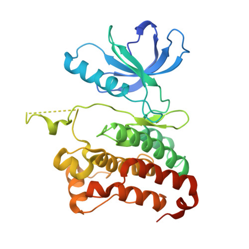
    Crystallographic studies of a range of 3-substituted anilinopyrimidine inhibitors of EphB4 have highlighted two alternative C-2 aniline conformations and this discovery has been exploited in the design of a highly potent series of 3,5-disubstituted anilinopyrimidines. The observed range of cellular activities has been rationalised on the basis of physicochemical and structural characteristics.


  • Organizational Affiliation

    AstraZeneca, Mereside, Alderley Park, Macclesfield, Cheshire SK10 4TG, UK.


Macromolecules
Find similar proteins by:  (by identity cutoff)  |  3D Structure
Entity ID: 1
MoleculeChains Sequence LengthOrganismDetailsImage
EPHRIN TYPE-B RECEPTOR 4302Homo sapiensMutation(s): 1 
EC: 2.7.10.1
UniProt & NIH Common Fund Data Resources
Find proteins for P54760 (Homo sapiens)
Explore P54760 
Go to UniProtKB:  P54760
PHAROS:  P54760
GTEx:  ENSG00000196411 
Entity Groups  
Sequence Clusters30% Identity50% Identity70% Identity90% Identity95% Identity100% Identity
UniProt GroupP54760
Sequence Annotations
Expand
  • Reference Sequence
Small Molecules
Binding Affinity Annotations 
IDSourceBinding Affinity
7X8 PDBBind:  2VX1 IC50: 220 (nM) from 1 assay(s)
BindingDB:  2VX1 IC50: min: 220, max: 900 (nM) from 2 assay(s)
Binding MOAD:  2VX1 IC50: 220 (nM) from 1 assay(s)
Experimental Data & Validation

Experimental Data

  • Method: X-RAY DIFFRACTION
  • Resolution: 1.65 Å
  • R-Value Free: 0.210 
  • R-Value Work: 0.171 
  • R-Value Observed: 0.173 
  • Space Group: P 1 21 1
Unit Cell:
Length ( Å )Angle ( ˚ )
a = 46.116α = 90
b = 53.537β = 110.9
c = 61.505γ = 90
Software Package:
Software NamePurpose
REFMACrefinement
CrystalCleardata reduction
CrystalCleardata scaling
AMoREphasing

Structure Validation

View Full Validation Report


Ligand Structure Quality Assessment 


Entry History 

Deposition Data

Revision History  (Full details and data files)

  • Version 1.0: 2008-10-28
    Type: Initial release
  • Version 1.1: 2011-05-08
    Changes: Version format compliance
  • Version 1.2: 2011-07-13
    Changes: Version format compliance
  • Version 1.3: 2017-06-28
    Changes: Data collection
  • Version 1.4: 2019-04-24
    Changes: Data collection, Source and taxonomy
  • Version 1.5: 2019-05-08
    Changes: Data collection, Experimental preparation
  • Version 1.6: 2023-12-13
    Changes: Data collection, Database references, Derived calculations, Other, Refinement description