2VST

hPPARgamma Ligand binding domain in complex with 13-(S)-HODE


Experimental Data Snapshot

  • Method: X-RAY DIFFRACTION
  • Resolution: 2.35 Å
  • R-Value Free: 0.274 
  • R-Value Work: 0.234 
  • R-Value Observed: 0.234 

wwPDB Validation   3D Report Full Report


Ligand Structure Quality Assessment 


This is version 1.2 of the entry. See complete history


Literature

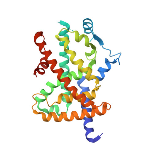
Structural Basis for the Activation of Pparg by Oxidised Fatty Acids

Itoh, T.Fairall, L.Amin, K.Inaba, Y.Szanto, A.Balint, B.L.Nagy, L.Yamamoto, K.Schwabe, J.W.R.

(2008) Nat Struct Mol Biol 15: 924

  • DOI: https://doi.org/10.1038/nsmb.1474
  • Primary Citation of Related Structures:  
    2VSR, 2VST, 2VV0, 2VV1, 2VV2, 2VV3, 2VV4

  • PubMed Abstract: 

    The nuclear receptor peroxisome proliferator-activated receptor-gamma (PPARgamma) has important roles in adipogenesis and immune response as well as roles in both lipid and carbohydrate metabolism. Although synthetic agonists for PPARgamma are widely used as insulin sensitizers, the identity of the natural ligand(s) for PPARgamma is still not clear. Suggested natural ligands include 15-deoxy-delta12,14-prostaglandin J2 and oxidized fatty acids such as 9-HODE and 13-HODE. Crystal structures of PPARgamma have revealed the mode of recognition for synthetic compounds. Here we report structures of PPARgamma bound to oxidized fatty acids that are likely to be natural ligands for this receptor. These structures reveal that the receptor can (i) simultaneously bind two fatty acids and (ii) couple covalently with conjugated oxo fatty acids. Thermal stability and gene expression analyses suggest that such covalent ligands are particularly effective activators of PPARgamma and thus may serve as potent and biologically relevant ligands.


  • Organizational Affiliation

    Henry Wellcome Laboratories of Structural Biology, Department of Biochemistry, University of Leicester, Lancaster Road, Leicester LE1 9HN, UK.


Macromolecules
Find similar proteins by:  (by identity cutoff)  |  3D Structure
Entity ID: 1
MoleculeChains Sequence LengthOrganismDetailsImage
PEROXISOME PROLIFERATOR-ACTIVATED RECEPTOR GAMMA
A, B
276Homo sapiensMutation(s): 0 
UniProt & NIH Common Fund Data Resources
Find proteins for P37231 (Homo sapiens)
Explore P37231 
Go to UniProtKB:  P37231
PHAROS:  P37231
GTEx:  ENSG00000132170 
Entity Groups  
Sequence Clusters30% Identity50% Identity70% Identity90% Identity95% Identity100% Identity
UniProt GroupP37231
Sequence Annotations
Expand
  • Reference Sequence
Small Molecules
Ligands 1 Unique
IDChains Name / Formula / InChI Key2D Diagram3D Interactions
243
Query on 243

Download Ideal Coordinates CCD File 
C [auth A],
D [auth B]
(9Z,11E,13S)-13-hydroxyoctadeca-9,11-dienoic acid
C18 H32 O3
HNICUWMFWZBIFP-IRQZEAMPSA-N
Experimental Data & Validation

Experimental Data

  • Method: X-RAY DIFFRACTION
  • Resolution: 2.35 Å
  • R-Value Free: 0.274 
  • R-Value Work: 0.234 
  • R-Value Observed: 0.234 
  • Space Group: C 1 2 1
Unit Cell:
Length ( Å )Angle ( ˚ )
a = 93.197α = 90
b = 61.37β = 102.73
c = 118.552γ = 90
Software Package:
Software NamePurpose
CNSrefinement
MOSFLMdata reduction
SCALAdata scaling
PHASERphasing

Structure Validation

View Full Validation Report


Ligand Structure Quality Assessment 


Entry History 

Deposition Data

Revision History  (Full details and data files)

  • Version 1.0: 2008-08-19
    Type: Initial release
  • Version 1.1: 2012-06-06
    Changes: Database references, Derived calculations, Non-polymer description, Other, Refinement description, Source and taxonomy, Version format compliance
  • Version 1.2: 2023-12-13
    Changes: Data collection, Database references, Derived calculations, Other, Refinement description