2VNJ

X-RAY STRUCTURE OF THE FERREDOXIN-NADP(H) REDUCTASE FROM RHODOBACTER CAPSULATUS IN COMPLEX WITH NADP. FORM I AT 2.13 ANGSTROMS RESOLUTION


Experimental Data Snapshot

  • Method: X-RAY DIFFRACTION
  • Resolution: 2.13 Å
  • R-Value Free: 0.271 
  • R-Value Work: 0.237 
  • R-Value Observed: 0.237 

wwPDB Validation   3D Report Full Report


Ligand Structure Quality Assessment 


This is version 1.4 of the entry. See complete history


Literature

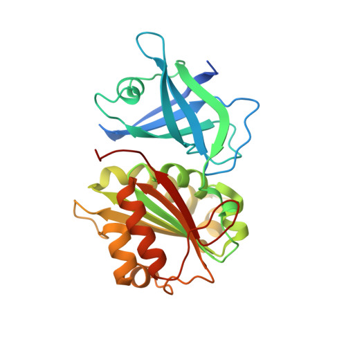
Coenzyme Binding and Hydride Transfer in Rhodobacter Capsulatus Ferredoxin/Flavodoxin Nadp(H) Oxidoreductase.

Bortolotti, A.Perez-Dorado, I.Goni, G.Medina, M.Hermoso, J.A.Carrillo, N.Cortez, N.

(2009) Biochim Biophys Acta 1794: 199

  • DOI: https://doi.org/10.1016/j.bbapap.2008.09.013
  • Primary Citation of Related Structures:  
    2VNH, 2VNI, 2VNJ, 2VNK

  • PubMed Abstract: 

    Ferredoxin-NADP(H) reductases catalyse the reversible hydride/electron exchange between NADP(H) and ferredoxin/flavodoxin, comprising a structurally defined family of flavoenzymes with two distinct subclasses. Those present in Gram-negative bacteria (FPRs) display turnover numbers of 1-5 s(-1) while the homologues of cyanobacteria and plants (FNRs) developed a 100-fold activity increase. We investigated nucleotide interactions and hydride transfer in Rhodobacter capsulatus FPR comparing them to those reported for FNRs. NADP(H) binding proceeds as in FNRs with stacking of the nicotinamide on the flavin, which resulted in formation of charge-transfer complexes prior to hydride exchange. The affinity of FPR for both NADP(H) and 2'-P-AMP was 100-fold lower than that of FNRs. The crystal structure of FPR in complex with 2'-P-AMP and NADP(+) allowed modelling of the adenosine ring system bound to the protein, whereas the nicotinamide portion was either not visible or protruding toward solvent in different obtained crystals. Stabilising contacts with the active site residues are different in the two reductase classes. We conclude that evolution to higher activities in FNRs was partially favoured by modification of NADP(H) binding in the initial complexes through changes in the active site residues involved in stabilisation of the adenosine portion of the nucleotide and in the mobile C-terminus of FPR.


  • Organizational Affiliation

    Instituto de Biología Molecular y Celular de Rosario, Universidad Nacional de Rosario, Suipacha 531, S2002LRK Rosario, Argentina.


Macromolecules
Find similar proteins by:  (by identity cutoff)  |  3D Structure
Entity ID: 1
MoleculeChains Sequence LengthOrganismDetailsImage
NADPH\:FERREDOXIN REDUCTASE272Rhodobacter capsulatusMutation(s): 0 
EC: 1.18.1.2
UniProt
Find proteins for Q9L6V3 (Rhodobacter capsulatus)
Explore Q9L6V3 
Go to UniProtKB:  Q9L6V3
Entity Groups  
Sequence Clusters30% Identity50% Identity70% Identity90% Identity95% Identity100% Identity
UniProt GroupQ9L6V3
Sequence Annotations
Expand
  • Reference Sequence
Small Molecules
Ligands 3 Unique
IDChains Name / Formula / InChI Key2D Diagram3D Interactions
FAD
Query on FAD

Download Ideal Coordinates CCD File 
B [auth A]FLAVIN-ADENINE DINUCLEOTIDE
C27 H33 N9 O15 P2
VWWQXMAJTJZDQX-UYBVJOGSSA-N
NAP
Query on NAP

Download Ideal Coordinates CCD File 
C [auth A]NADP NICOTINAMIDE-ADENINE-DINUCLEOTIDE PHOSPHATE
C21 H28 N7 O17 P3
XJLXINKUBYWONI-NNYOXOHSSA-N
HTG
Query on HTG

Download Ideal Coordinates CCD File 
D [auth A]heptyl 1-thio-beta-D-glucopyranoside
C13 H26 O5 S
HPEGNLMTTNTJSP-LBELIVKGSA-N
Binding Affinity Annotations 
IDSourceBinding Affinity
NAP Binding MOAD:  2VNJ Kd: 2.22e+5 (nM) from 1 assay(s)
Experimental Data & Validation

Experimental Data

  • Method: X-RAY DIFFRACTION
  • Resolution: 2.13 Å
  • R-Value Free: 0.271 
  • R-Value Work: 0.237 
  • R-Value Observed: 0.237 
  • Space Group: P 31 2 1
Unit Cell:
Length ( Å )Angle ( ˚ )
a = 68.72α = 90
b = 68.72β = 90
c = 128.15γ = 120
Software Package:
Software NamePurpose
CNSrefinement
MOSFLMdata reduction
SCALAdata scaling
MOLREPphasing

Structure Validation

View Full Validation Report


Ligand Structure Quality Assessment 


Entry History 

Deposition Data

Revision History  (Full details and data files)

  • Version 1.0: 2008-11-11
    Type: Initial release
  • Version 1.1: 2011-05-08
    Changes: Version format compliance
  • Version 1.2: 2011-07-13
    Changes: Version format compliance
  • Version 1.3: 2020-07-29
    Type: Remediation
    Reason: Carbohydrate remediation
    Changes: Derived calculations, Other, Structure summary
  • Version 1.4: 2023-12-13
    Changes: Data collection, Database references, Refinement description, Structure summary