2VII

PspF1-275-Mg-AMP


Experimental Data Snapshot

  • Method: X-RAY DIFFRACTION
  • Resolution: 2.85 Å
  • R-Value Free: 0.241 
  • R-Value Work: 0.186 
  • R-Value Observed: 0.186 

wwPDB Validation   3D Report Full Report


Ligand Structure Quality Assessment 


This is version 1.3 of the entry. See complete history


Literature

Trapping of a Transcription Complex Using a New Nucleotide Analogue: AMP Aluminium Fluoride

Joly, N.Rappas, M.Buck, M.Zhang, X.

(2008) J Mol Biol 375: 1206

  • DOI: https://doi.org/10.1016/j.jmb.2007.11.050
  • Primary Citation of Related Structures:  
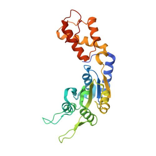
    2VII

  • PubMed Abstract: 

    Mechanochemical proteins rely on ATP hydrolysis to establish the different functional states required for their biological output. Studying the transient functional intermediate states these proteins adopt as they progress through the ATP hydrolysis cycle is key to understanding the molecular basis of their mechanism. Many of these intermediates have been successfully 'trapped' and functionally characterised using ATP analogues. Here, we present a new nucleotide analogue, AMP-AlF(x), which traps PspF, a bacterial enhancer binding protein, in a stable complex with the sigma(54)-RNA polymerase holoenzyme. The crystal structure of AMP-AlF(x)*PspF(1-275) provides new information on protein-nucleotide interactions and suggests that the beta and gamma phosphates are more important than the alpha phosphate in terms of sensing nucleotide bound states. In addition, functional data obtained with AMP-AlF(x) establish distinct roles for the conserved catalytic AAA(+) (ATPases associated with various cellular activities) residues, suggesting that AMP-AlF(x) is a powerful new tool to study AAA(+) protein family members and, more generally, Walker motif ATPases.


  • Organizational Affiliation

    Division of Biology, Sir Alexander Fleming Building, Imperial College London, Exhibition Road, South Kensington, London SW7 2AZ, UK.


Macromolecules
Find similar proteins by:  (by identity cutoff)  |  3D Structure
Entity ID: 1
MoleculeChains Sequence LengthOrganismDetailsImage
PSP OPERON TRANSCRIPTIONAL ACTIVATOR259Escherichia coli K-12Mutation(s): 0 
UniProt
Find proteins for P37344 (Escherichia coli (strain K12))
Explore P37344 
Go to UniProtKB:  P37344
Entity Groups  
Sequence Clusters30% Identity50% Identity70% Identity90% Identity95% Identity100% Identity
UniProt GroupP37344
Sequence Annotations
Expand
  • Reference Sequence
Small Molecules
Ligands 2 Unique
IDChains Name / Formula / InChI Key2D Diagram3D Interactions
AMP
Query on AMP

Download Ideal Coordinates CCD File 
B [auth A]ADENOSINE MONOPHOSPHATE
C10 H14 N5 O7 P
UDMBCSSLTHHNCD-KQYNXXCUSA-N
MG
Query on MG

Download Ideal Coordinates CCD File 
C [auth A]MAGNESIUM ION
Mg
JLVVSXFLKOJNIY-UHFFFAOYSA-N
Experimental Data & Validation

Experimental Data

  • Method: X-RAY DIFFRACTION
  • Resolution: 2.85 Å
  • R-Value Free: 0.241 
  • R-Value Work: 0.186 
  • R-Value Observed: 0.186 
  • Space Group: P 65
Unit Cell:
Length ( Å )Angle ( ˚ )
a = 113.607α = 90
b = 113.607β = 90
c = 39.358γ = 120
Software Package:
Software NamePurpose
CNSrefinement
MOSFLMdata reduction
SCALAdata scaling
PHASERphasing

Structure Validation

View Full Validation Report


Ligand Structure Quality Assessment 


Entry History 

Deposition Data

Revision History  (Full details and data files)

  • Version 1.0: 2008-01-22
    Type: Initial release
  • Version 1.1: 2011-05-08
    Changes: Version format compliance
  • Version 1.2: 2011-07-13
    Changes: Version format compliance
  • Version 1.3: 2023-12-13
    Changes: Data collection, Database references, Derived calculations, Other, Refinement description