2VHB

AZIDE ADDUCT OF THE BACTERIAL HEMOGLOBIN FROM VITREOSCILLA STERCORARIA


Experimental Data Snapshot

  • Method: X-RAY DIFFRACTION
  • Resolution: 1.76 Å
  • R-Value Work: 0.197 

wwPDB Validation   3D Report Full Report


Ligand Structure Quality Assessment 


This is version 1.3 of the entry. See complete history


Literature

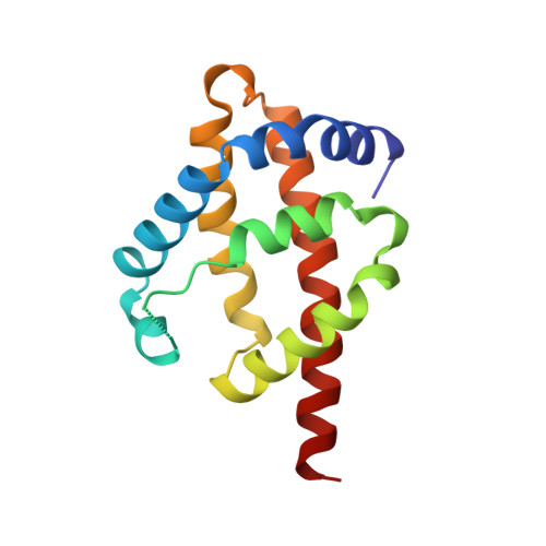
Unusual structure of the oxygen-binding site in the dimeric bacterial hemoglobin from Vitreoscilla sp.

Tarricone, C.Galizzi, A.Coda, A.Ascenzi, P.Bolognesi, M.

(1997) Structure 5: 497-507

  • DOI: https://doi.org/10.1016/s0969-2126(97)00206-2
  • Primary Citation of Related Structures:  
    1VHB, 2VHB

  • PubMed Abstract: 

    The first hemoglobin identified in bacteria was isolated from Vitreoscilla stercoraria (VtHb) as a homodimeric species. The wild-type protein has been reported to display medium oxygen affinity and cooperative ligand-binding properties. Moreover, VtHb can support aerobic growth in Escherichia coli with impaired terminal oxidase function. This ability of VtHb to improve the growth properties of E. coli has important applications in fermentation technology, assisting the overexpression of recombinant proteins and antibiotics. Oxygen binding heme domains have been identified in chimeric proteins from bacteria and yeast, where they are covalently linked to FAD- and NAD(P)H-binding domains. We investigate here the fold, the distal heme site structure and the quaternary assembly of a bacterial hemoglobin which does not bear the typical flavohemoglobin domain organization.


  • Organizational Affiliation

    Dipartimento di Genetica e Microbiologia, Università di Pavia, Via Abbiategrasso 207, 27100, Pavia, Italy.


Macromolecules
Find similar proteins by:  (by identity cutoff)  |  3D Structure
Entity ID: 1
MoleculeChains Sequence LengthOrganismDetailsImage
HEMOGLOBIN
A, B
146Vitreoscilla stercorariaMutation(s): 0 
Gene Names: VGB
UniProt
Find proteins for P04252 (Vitreoscilla stercoraria)
Explore P04252 
Go to UniProtKB:  P04252
Entity Groups  
Sequence Clusters30% Identity50% Identity70% Identity90% Identity95% Identity100% Identity
UniProt GroupP04252
Sequence Annotations
Expand
  • Reference Sequence
Experimental Data & Validation

Experimental Data

  • Method: X-RAY DIFFRACTION
  • Resolution: 1.76 Å
  • R-Value Work: 0.197 
  • Space Group: P 1 21 1
Unit Cell:
Length ( Å )Angle ( ˚ )
a = 62.72α = 90
b = 40.19β = 106.55
c = 62.94γ = 90
Software Package:
Software NamePurpose
TNTrefinement
DENZOdata reduction
SCALEPACKdata scaling
TNTphasing

Structure Validation

View Full Validation Report


Ligand Structure Quality Assessment 


Entry History 

Deposition Data

Revision History  (Full details and data files)

  • Version 1.0: 1998-02-25
    Type: Initial release
  • Version 1.1: 2008-03-25
    Changes: Version format compliance
  • Version 1.2: 2011-07-13
    Changes: Version format compliance
  • Version 1.3: 2024-02-21
    Changes: Data collection, Database references, Derived calculations