2VEB

High resolution structure of protoglobin from Methanosarcina acetivorans C2A


Experimental Data Snapshot

  • Method: X-RAY DIFFRACTION
  • Resolution: 1.30 Å
  • R-Value Free: 0.190 
  • R-Value Work: 0.160 
  • R-Value Observed: 0.161 

wwPDB Validation   3D Report Full Report


Ligand Structure Quality Assessment 


This is version 1.1 of the entry. See complete history


Literature

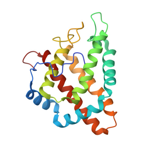
Archaeal Protoglobin Structure Indicates New Ligand Diffusion Paths and Modulation of Haem-Reactivity.

Nardini, M.Pesce, A.Thijs, L.Saito, J.A.Dewilde, S.Alam, M.Ascenzi, P.Coletta, M.Ciaccio, C.Moens, L.Bolognesi, M.

(2008) EMBO Rep 9: 157

  • DOI: https://doi.org/10.1038/sj.embor.7401153
  • Primary Citation of Related Structures:  
    2VEB, 2VEE

  • PubMed Abstract: 

    The structural adaptability of the globin fold has been highlighted by the recent discovery of the 2-on-2 haemoglobins, of neuroglobin and cytoglobin. Protoglobin from Methanosarcina acetivorans C2A-a strictly anaerobic methanogenic Archaea-is, to the best of our knowledge, the latest entry adding new variability and functional complexity to the haemoglobin (Hb) superfamily. Here, we report the 1.3 A crystal structure of oxygenated M. acetivorans protoglobin, together with the first insight into its ligand-binding properties. We show that, contrary to all known globins, protoglobin-specific loops and an amino-terminal extension completely bury the haem within the protein matrix. Access of O(2), CO and NO to the haem is granted by the protoglobin-specific apolar tunnels reaching the haem distal site from locations at the B/G and B/E helix interfaces. Functionally, M. acetivorans dimeric protoglobin shows a selectivity ratio for O(2)/CO binding to the haem that favours O(2) ligation and anticooperativity in ligand binding. Both properties are exceptional within the Hb superfamily.


  • Organizational Affiliation

    Department of Biomolecular Sciences and Biotechnology, CNR-INFM, University of Milano, Via Celoria 26, Milano 20133, Italy.


Macromolecules
Find similar proteins by:  (by identity cutoff)  |  3D Structure
Entity ID: 1
MoleculeChains Sequence LengthOrganismDetailsImage
PROTOGLOBIN195Methanosarcina acetivoransMutation(s): 1 
UniProt
Find proteins for Q8TLY9 (Methanosarcina acetivorans (strain ATCC 35395 / DSM 2834 / JCM 12185 / C2A))
Explore Q8TLY9 
Go to UniProtKB:  Q8TLY9
Entity Groups  
Sequence Clusters30% Identity50% Identity70% Identity90% Identity95% Identity100% Identity
UniProt GroupQ8TLY9
Sequence Annotations
Expand
  • Reference Sequence
Experimental Data & Validation

Experimental Data

  • Method: X-RAY DIFFRACTION
  • Resolution: 1.30 Å
  • R-Value Free: 0.190 
  • R-Value Work: 0.160 
  • R-Value Observed: 0.161 
  • Space Group: C 1 2 1
Unit Cell:
Length ( Å )Angle ( ˚ )
a = 80.059α = 90
b = 49.271β = 92.88
c = 51.513γ = 90
Software Package:
Software NamePurpose
REFMACrefinement
MOSFLMdata reduction
SCALAdata scaling
SOLVEphasing

Structure Validation

View Full Validation Report


Ligand Structure Quality Assessment 


Entry History 

Deposition Data

Revision History  (Full details and data files)

  • Version 1.0: 2008-01-22
    Type: Initial release
  • Version 1.1: 2012-08-22
    Changes: Advisory, Derived calculations, Non-polymer description, Other, Version format compliance