2V6C

Crystal structure of ErbB3 binding protein 1 (Ebp1)


Experimental Data Snapshot

  • Method: X-RAY DIFFRACTION
  • Resolution: 2.50 Å
  • R-Value Free: 0.213 
  • R-Value Work: 0.182 
  • R-Value Observed: 0.182 

wwPDB Validation   3D Report Full Report


This is version 1.4 of the entry. See complete history


Literature

Structural Insights Into the Transcriptional and Translational Roles of Ebp1

Monie, T.P.Perrin, A.J.Birtley, J.R.Sweeney, T.R.Karakasiliotis, I.Chaudry, Y.Roberts, L.O.Matthews, S.Goodfellow, I.G.Curry, S.

(2007) EMBO J 26: 3936

  • DOI: https://doi.org/10.1038/sj.emboj.7601817
  • Primary Citation of Related Structures:  
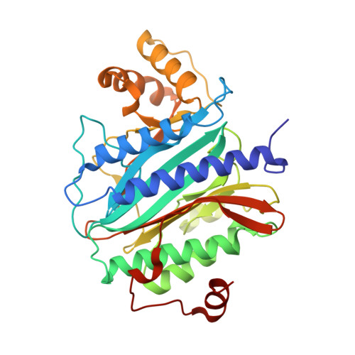
    2V6C

  • PubMed Abstract: 

    The ErbB3-binding protein 1 (Ebp1) is an important regulator of transcription, affecting eukaryotic cell growth, proliferation, differentiation and survival. Ebp1 can also affect translation and cooperates with the polypyrimidine tract-binding protein (PTB) to stimulate the activity of the internal ribosome entry site (IRES) of foot-and-mouth disease virus (FMDV). We report here the crystal structure of murine Ebp1 (p48 isoform), providing the first glimpse of the architecture of this versatile regulator. The structure reveals a core domain that is homologous to methionine aminopeptidases, coupled to a C-terminal extension that contains important motifs for binding proteins and RNA. It sheds new light on the conformational differences between the p42 and p48 isoforms of Ebp1, the disposition of the key protein-interacting motif ((354)LKALL(358)) and the RNA-binding activity of Ebp1. We show that the primary RNA-binding site is formed by a Lys-rich motif in the C terminus and mediates the interaction with the FMDV IRES. We also demonstrate a specific functional requirement for Ebp1 in FMDV IRES-directed translation that is independent of a direct interaction with PTB.


  • Organizational Affiliation

    Division of Cell and Molecular Biology, Imperial College, South Kensington Campus, London, UK.


Macromolecules
Find similar proteins by:  (by identity cutoff)  |  3D Structure
Entity ID: 1
MoleculeChains Sequence LengthOrganismDetailsImage
PROLIFERATION-ASSOCIATED PROTEIN 2G4353Mus musculusMutation(s): 0 
UniProt & NIH Common Fund Data Resources
Find proteins for P50580 (Mus musculus)
Explore P50580 
Go to UniProtKB:  P50580
IMPC:  MGI:894684
Entity Groups  
Sequence Clusters30% Identity50% Identity70% Identity90% Identity95% Identity100% Identity
UniProt GroupP50580
Sequence Annotations
Expand
  • Reference Sequence
Experimental Data & Validation

Experimental Data

  • Method: X-RAY DIFFRACTION
  • Resolution: 2.50 Å
  • R-Value Free: 0.213 
  • R-Value Work: 0.182 
  • R-Value Observed: 0.182 
  • Space Group: P 21 21 21
Unit Cell:
Length ( Å )Angle ( ˚ )
a = 49.147α = 90
b = 72.779β = 90
c = 115.624γ = 90
Software Package:
Software NamePurpose
CNSrefinement
MOSFLMdata reduction
SCALAdata scaling
PHASERphasing

Structure Validation

View Full Validation Report


Entry History 

Deposition Data

Revision History  (Full details and data files)

  • Version 1.0: 2007-08-21
    Type: Initial release
  • Version 1.1: 2011-05-08
    Changes: Version format compliance
  • Version 1.2: 2011-07-13
    Changes: Version format compliance
  • Version 1.3: 2019-07-24
    Changes: Data collection
  • Version 1.4: 2023-12-13
    Changes: Data collection, Database references, Other, Refinement description