2UVF

Structure of Yersinia enterocolitica Family 28 Exopolygalacturonase in Complex with Digalaturonic Acid


Experimental Data Snapshot

  • Method: X-RAY DIFFRACTION
  • Resolution: 2.10 Å
  • R-Value Free: 0.284 
  • R-Value Work: 0.232 
  • R-Value Observed: 0.234 

wwPDB Validation   3D Report Full Report


This is version 2.1 of the entry. See complete history


Literature

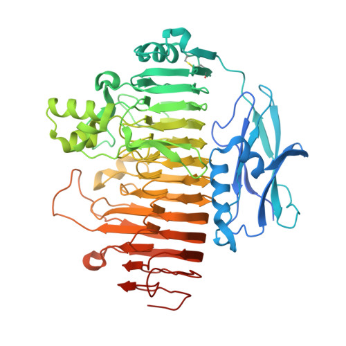
The Structural Basis for Exopolygalacturonase Activity in a Family 28 Glycoside Hydrolase.

Abbott, D.W.Boraston, A.B.

(2007) J Mol Biol 368: 1215

  • DOI: https://doi.org/10.1016/j.jmb.2007.02.083
  • Primary Citation of Related Structures:  
    2UVE, 2UVF

  • PubMed Abstract: 

    Family 28 glycoside hydrolases (polygalacturonases) are found in organisms across the plant, fungal and bacterial kingdoms, where they are central to diverse biological functions such as fruit ripening, biomass recycling and plant pathogenesis. The structures of several polygalacturonases have been reported; however, all of these enzymes utilize an endo-mode of digestion, which generates a spectrum of oligosaccharide products with varying degrees of polymerization. The structure of a complementary exo-acting polygalacturonase and an accompanying explanation of the molecular determinants for its specialized activity have been noticeably lacking. We present the structure of an exopolygalacturonase from Yersinia enterocolitica, YeGH28 in a native form (solved to 2.19 A resolution) and a digalacturonic acid product complex (solved to 2.10 A resolution). The activity of YeGH28 is due to inserted stretches of amino acid residues that transform the active site from the open-ended channel observed in the endopolygalacturonases to a closed pocket that restricts the enzyme to the exclusive attack of the non-reducing end of oligogalacturonide substrates. In addition, YeGH28 possesses a fused FN3 domain with unknown function, the first such structure described in pectin active enzymes.


  • Organizational Affiliation

    Biochemistry and Microbiology, University of Victoria, PO Box 3055 STN CSC, Victoria BC, Canada V8W 3P6.


Macromolecules
Find similar proteins by:  (by identity cutoff)  |  3D Structure
Entity ID: 1
MoleculeChains Sequence LengthOrganismDetailsImage
EXOPOLYGALACTURONASE
A, B
608Yersinia enterocoliticaMutation(s): 0 
UniProt
Find proteins for O68975 (Yersinia enterocolitica)
Explore O68975 
Go to UniProtKB:  O68975
Entity Groups  
Sequence Clusters30% Identity50% Identity70% Identity90% Identity95% Identity100% Identity
UniProt GroupO68975
Sequence Annotations
Expand
  • Reference Sequence
Oligosaccharides

Help

Entity ID: 2
MoleculeChains Length2D Diagram Glycosylation3D Interactions
alpha-D-galactopyranuronic acid-(1-4)-alpha-D-galactopyranuronic acid
C
2N/A
Glycosylation Resources
GlyTouCan:  G65215OP
GlyCosmos:  G65215OP
Small Molecules
Ligands 4 Unique
IDChains Name / Formula / InChI Key2D Diagram3D Interactions
PEG
Query on PEG

Download Ideal Coordinates CCD File 
JA [auth B],
KA [auth B]
DI(HYDROXYETHYL)ETHER
C4 H10 O3
MTHSVFCYNBDYFN-UHFFFAOYSA-N
SO4
Query on SO4

Download Ideal Coordinates CCD File 
D [auth A]
E [auth A]
F [auth A]
G [auth A]
H [auth A]
D [auth A],
E [auth A],
F [auth A],
G [auth A],
H [auth A],
I [auth A],
N [auth B],
O [auth B],
P [auth B],
Q [auth B],
R [auth B],
S [auth B],
T [auth B],
U [auth B],
V [auth B],
W [auth B],
X [auth B],
Y [auth B]
SULFATE ION
O4 S
QAOWNCQODCNURD-UHFFFAOYSA-L
ACT
Query on ACT

Download Ideal Coordinates CCD File 
FA [auth B],
GA [auth B],
HA [auth B],
IA [auth B]
ACETATE ION
C2 H3 O2
QTBSBXVTEAMEQO-UHFFFAOYSA-M
NI
Query on NI

Download Ideal Coordinates CCD File 
AA [auth B]
BA [auth B]
CA [auth B]
DA [auth B]
EA [auth B]
AA [auth B],
BA [auth B],
CA [auth B],
DA [auth B],
EA [auth B],
J [auth A],
K [auth A],
L [auth A],
M [auth A],
Z [auth B]
NICKEL (II) ION
Ni
VEQPNABPJHWNSG-UHFFFAOYSA-N
Experimental Data & Validation

Experimental Data

  • Method: X-RAY DIFFRACTION
  • Resolution: 2.10 Å
  • R-Value Free: 0.284 
  • R-Value Work: 0.232 
  • R-Value Observed: 0.234 
  • Space Group: P 1 21 1
Unit Cell:
Length ( Å )Angle ( ˚ )
a = 91.015α = 90
b = 79.659β = 103.9
c = 98.493γ = 90
Software Package:
Software NamePurpose
REFMACrefinement

Structure Validation

View Full Validation Report


Entry History 

Deposition Data

Revision History  (Full details and data files)

  • Version 1.0: 2007-05-08
    Type: Initial release
  • Version 1.1: 2011-05-08
    Changes: Version format compliance
  • Version 1.2: 2011-07-13
    Changes: Version format compliance
  • Version 2.0: 2020-07-29
    Type: Remediation
    Reason: Carbohydrate remediation
    Changes: Advisory, Atomic model, Data collection, Derived calculations, Non-polymer description, Other, Structure summary
  • Version 2.1: 2023-12-13
    Changes: Data collection, Database references, Refinement description, Structure summary