Structure and refinement of the oxidized P21 form of uteroglobin at 1.64 A resolution.
Bally, R., Delettre, J.(1989) J Mol Biol 206: 153-170
- PubMed: 2704039
- DOI: https://doi.org/10.1016/0022-2836(89)90530-5
- Primary Citation of Related Structures:
2UTG - PubMed Abstract:
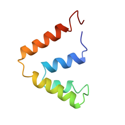
One of the monoclinic P21 forms of uteroglobin, a progesterone-binding protein secreted by the rabbit uterus, was crystallized and subjected to X-ray diffraction analysis at 1.64 A resolution. The analysis was refined to an R factor of 0.19 and the 1096 non-hydrogen atomic positions are known to an accuracy of about 0.18 A. The average isotropic temperature factor B was 10.4 A2. Uteroglobin is a dimer of two independent polypeptide chains of 70 residues linked by two disulfide bridges and related by a pseudo binary axis. Each monomer is folded into four alpha-helices. An oblong hydrophobic pocket is observed inside the dimer, and the possibility that it represents a progesterone-binding site is discussed. The present model includes 165 possible sites for water molecules, of which six are located in the hydrophobic pocket. Polar groups are involved in hydrogen bonding (intramolecular, intermolecular or with water molecules).
Organizational Affiliation:
Laboratoire de Minéralogie-Cristallographie associé au CNRS, Université Paris, France.