NMR structure, localization, and vesicle fusion of Chikungunya virus fusion peptide
Mohanram, H., Nip, A., Domadia, P.N., Bhunia, A., Bhattacharjya, S.(2012) Biochemistry 51: 7863-7872
- PubMed: 22978677
- DOI: https://doi.org/10.1021/bi300901f
- Primary Citation of Related Structures:
2RSW - PubMed Abstract:
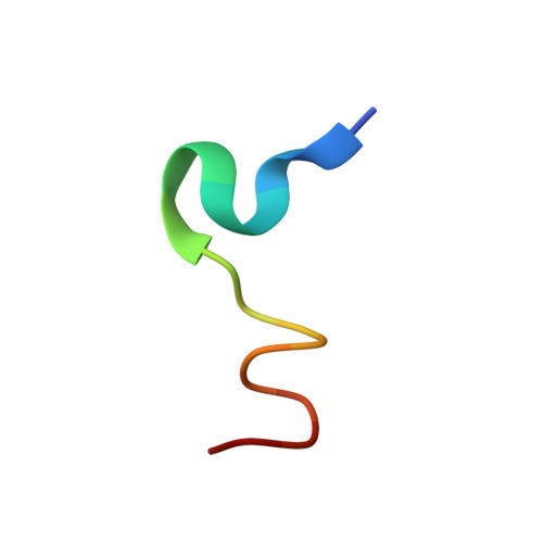
The virus-host cell fusion process is mediated by a membrane anchored viral fusion protein that inserts its hydrophobic fusion peptide into the plasma membrane of the host cell, initiating the fusion reaction. Therefore, fusion peptides are an important functional constituent of the fusion proteins of enveloped viruses. In this work, we characterize the fusion peptide or VT18 (V(84)YPFMWGGAYCFCDAENT(101)) of Chikungunya virus (CHIKV) using NMR and fluorescence spectroscopy in zwitterionic lipid environments. Our results demonstrate that the VT18 peptide is able to induce liposome fusions in a pH independent manner and interacts with the zwitterionic lipid vesicles. The NMR derived three-dimensional structure of VT18, in solution of dodecylphosphocholine (DPC) micelles, is typified by extended or β-type conformations for most of the residues, whereby residues M88-W89-G90-G91 adopt a type I β-turn conformation. Strikingly, the aromatic side chains of residues Y85, F87, Y93, and F95 in the VT18 structure are found to be well-packed forming an aromatic core. In particular, residue F87 is situated at the center of the aromatic core establishing a close proximity with other aromatic side chains. Further, the aromatic core residues are also involved in packing interactions with the side chains of residues M88, C94. Paramagnetic relaxation enhancement NMR, using spin labeled doxyl lipids, indicated that the aromatic core residues of VT18 are well inserted into the micelles, whereas the polar residues at the C-terminus may be surface localized. The atomic resolution structure and lipid interactions of CHIKV fusion peptide presented here will aid to uncover the fusion mechanism by the type II viral fusion proteins.
Organizational Affiliation:
School of Biological Sciences, Division of Structural Biology and Biochemistry, Nanyang Technological University, Singapore 637551.