Antimicrobial peptide RP-1 structure and interactions with anionic versus zwitterionic micelles.
Bourbigot, S., Dodd, E., Horwood, C., Cumby, N., Fardy, L., Welch, W.H., Ramjan, Z., Sharma, S., Waring, A.J., Yeaman, M.R., Booth, V.(2009) Biopolymers 91: 1-13
- PubMed: 18712851
- DOI: https://doi.org/10.1002/bip.21071
- Primary Citation of Related Structures:
2RLG, 2RLH - PubMed Abstract:
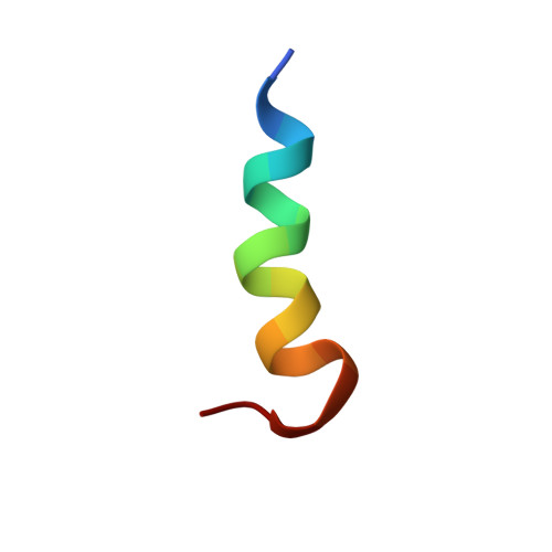
Topologically, platelet factor-4 kinocidins consist of distinct N-terminal extended, C-terminal helical, and interposing gamma-core structural domains. The C-terminal alpha-helices autonomously confer direct microbicidal activity, and the synthetic antimicrobial peptide RP-1 is modeled upon these domains. In this study, the structure of RP-1 was assessed using several complementary techniques. The high-resolution structure of RP-1 was determined by NMR in anionic sodium dodecyl sulfate (SDS) and zwitterionic dodecylphosphocholine (DPC) micelles, which approximate prokaryotic and eukaryotic membranes, respectively. NMR data indicate the peptide assumes an amphipathic alpha-helical backbone conformation in both micelle environments. However, small differences were observed in the side-chain orientations of lysine, tyrosine, and phenylalanine residues in SDS versus DPC environments. NMR experiments with a paramagnetic probe indicated differences in positioning of the peptide within the two micelle types. Molecular dynamics (MD) simulations of the peptide in both micelle types were also performed to add insight into the peptide/micelle interactions and to assess the validity of this technique to predict the structure of peptides in complex with micelles. MD independently predicted RP-1 to interact only peripherally with the DPC micelle, leaving its spherical shape intact. In contrast, RP-1 entered deeply into and significantly distorted the SDS micelle. Overall, the experimental and MD results support a preferential specificity of RP-1 for anionic membranes over zwitterionic membranes. This specificity likely derives from differences in RP-1 interaction with distinct lipid systems, including subtle differences in side chain orientations, rather than gross changes in RP-1 structure in the two lipid environments.
Organizational Affiliation:
Department of Biochemistry, Memorial University of Newfoundland, St. John's, Newfoundland A1B 3X9, Canada.