2RHP

The Thrombospondin-1 Polymorphism Asn700Ser Associated with Cornoary Artery Disease Causes Local and Long-Ranging Changes in Protein Structure


Experimental Data Snapshot

  • Method: X-RAY DIFFRACTION
  • Resolution: 2.90 Å
  • R-Value Free: 0.277 
  • R-Value Work: 0.227 
  • R-Value Observed: 0.229 

wwPDB Validation   3D Report Full Report


Ligand Structure Quality Assessment 


This is version 2.2 of the entry. See complete history


Literature

Influences of the N700S Thrombospondin-1 Polymorphism on Protein Structure and Stability.

Carlson, C.B.Liu, Y.Keck, J.L.Mosher, D.F.

(2008) J Biol Chem 283: 20069-20076

  • DOI: https://doi.org/10.1074/jbc.M800223200
  • Primary Citation of Related Structures:  
    2RHP

  • PubMed Abstract: 

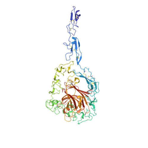
    Thrombospondins (THBSs) are multimodular, secreted proteins characterized by a signature domain comprising a unique set of 13 calcium-binding repeats flanked by epidermal growth factor (EGF)-like and lectin-like modules. A polymorphism that changes a conserved Asn to Ser at residue 700 in the most N-terminal calcium-binding repeat of THBS-1 (repeat 1C) is found in 8-10% of European populations and has been linked to increased risk of premature coronary artery disease. The Ser substitution leads to altered stability in the EGF-like and wire modules of the THBS-1 signature domain as assessed by differential scanning calorimetry carried out in 2 mm or 200 mum calcium. Studies of the melting profiles of the THBS-2 signature domain proteins with Asn or Ser at position 702 (homologous to 700 in THBS-1) revealed that the impact of the Ser allele is similar in both THBS-1 and THBS-2. Structure determination of the Ser(702) THBS-2 variant in 2 mm calcium showed that repeat 1C contains two bound calcium ions as in the crystal of the Asn(702) protein, including the ion that is coordinated by Asn(702), and is associated with changes in conformation of repeat 1C and the adjacent EGF-like modules. The Ser substitution leads to the decreased ability of soluble THBS-2 signature domain protein to bind 4B6.13, a conformation-sensitive monoclonal antibody that recognizes an epitope in repeat 1C. These results indicate that although THBS harboring the Ser allele binds a full complement of calcium ions, repeat 1C is altered, leading to destabilization of surrounding structures.


  • Organizational Affiliation

    Department of Medicine and Biomolecular Chemistry, University of Wisconsin, Madison, WI 53706, USA.


Macromolecules
Find similar proteins by:  (by identity cutoff)  |  3D Structure
Entity ID: 1
MoleculeChains Sequence LengthOrganismDetailsImage
Thrombospondin-2622Homo sapiensMutation(s): 1 
Gene Names: THBS2TSP2
UniProt & NIH Common Fund Data Resources
Find proteins for P35442 (Homo sapiens)
Explore P35442 
Go to UniProtKB:  P35442
PHAROS:  P35442
GTEx:  ENSG00000186340 
Entity Groups  
Sequence Clusters30% Identity50% Identity70% Identity90% Identity95% Identity100% Identity
UniProt GroupP35442
Sequence Annotations
Expand
  • Reference Sequence
Oligosaccharides

Help

Entity ID: 2
MoleculeChains Length2D Diagram Glycosylation3D Interactions
2-acetamido-2-deoxy-beta-D-glucopyranose-(1-4)-2-acetamido-2-deoxy-beta-D-glucopyranose
B
2N-Glycosylation
Glycosylation Resources
GlyTouCan:  G42666HT
GlyCosmos:  G42666HT
GlyGen:  G42666HT
Small Molecules
Ligands 2 Unique
IDChains Name / Formula / InChI Key2D Diagram3D Interactions
NAG
Query on NAG

Download Ideal Coordinates CCD File 
C [auth A],
D [auth A]
2-acetamido-2-deoxy-beta-D-glucopyranose
C8 H15 N O6
OVRNDRQMDRJTHS-FMDGEEDCSA-N
CA
Query on CA

Download Ideal Coordinates CCD File 
AA [auth A]
BA [auth A]
CA [auth A]
DA [auth A]
E [auth A]
AA [auth A],
BA [auth A],
CA [auth A],
DA [auth A],
E [auth A],
EA [auth A],
F [auth A],
FA [auth A],
G [auth A],
GA [auth A],
H [auth A],
HA [auth A],
I [auth A],
J [auth A],
K [auth A],
L [auth A],
M [auth A],
N [auth A],
O [auth A],
P [auth A],
Q [auth A],
R [auth A],
S [auth A],
T [auth A],
U [auth A],
V [auth A],
W [auth A],
X [auth A],
Y [auth A],
Z [auth A]
CALCIUM ION
Ca
BHPQYMZQTOCNFJ-UHFFFAOYSA-N
Experimental Data & Validation

Experimental Data

  • Method: X-RAY DIFFRACTION
  • Resolution: 2.90 Å
  • R-Value Free: 0.277 
  • R-Value Work: 0.227 
  • R-Value Observed: 0.229 
  • Space Group: I 2 2 2
Unit Cell:
Length ( Å )Angle ( ˚ )
a = 93.892α = 90
b = 122.65β = 90
c = 155.381γ = 90
Software Package:
Software NamePurpose
AMoREphasing
REFMACrefinement
PDB_EXTRACTdata extraction
HKL-2000data collection
DENZOdata reduction
SCALEPACKdata scaling
HKL-2000data scaling

Structure Validation

View Full Validation Report


Ligand Structure Quality Assessment 


Entry History 

Deposition Data

Revision History  (Full details and data files)

  • Version 1.0: 2008-05-27
    Type: Initial release
  • Version 1.1: 2011-07-13
    Changes: Non-polymer description, Version format compliance
  • Version 1.2: 2017-10-11
    Changes: Data collection, Refinement description
  • Version 2.0: 2020-07-29
    Type: Remediation
    Reason: Carbohydrate remediation
    Changes: Atomic model, Data collection, Derived calculations, Structure summary
  • Version 2.1: 2021-10-20
    Changes: Database references, Structure summary
  • Version 2.2: 2023-08-30
    Changes: Data collection, Refinement description