


Crystal structure of CBS domain, NE2398.
Dong, A., Xu, X., Korniyenko, A., Yakunin, A., Zheng, H., Walker, J.R., Edwards, A.M., Joachimiak, A., Savchenko, A.To be published.
Experimental Data Snapshot
Entity ID: 1 | |||||
|---|---|---|---|---|---|
| Molecule | Chains | Sequence Length | Organism | Details | Image |
| CBS domain | 135 | Nitrosomonas europaea ATCC 19718 | Mutation(s): 0 Gene Names: NE2398 | 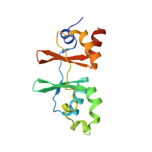 | |
UniProt | |||||
Find proteins for Q82SE2 (Nitrosomonas europaea (strain ATCC 19718 / CIP 103999 / KCTC 2705 / NBRC 14298)) Explore Q82SE2 Go to UniProtKB: Q82SE2 | |||||
Entity Groups | |||||
| Sequence Clusters | 30% Identity50% Identity70% Identity90% Identity95% Identity100% Identity | ||||
| UniProt Group | Q82SE2 | ||||
Sequence AnnotationsExpand | |||||
| |||||
| Ligands 2 Unique | |||||
|---|---|---|---|---|---|
| ID | Chains | Name / Formula / InChI Key | 2D Diagram | 3D Interactions | |
| NAD Query on NAD | GA [auth C], M [auth A], S [auth B], UA [auth D] | NICOTINAMIDE-ADENINE-DINUCLEOTIDE C21 H27 N7 O14 P2 BAWFJGJZGIEFAR-NNYOXOHSSA-N |  | ||
| BR Query on BR | AA [auth C] BA [auth C] CA [auth C] DA [auth C] E [auth A] | BROMIDE ION Br CPELXLSAUQHCOX-UHFFFAOYSA-M |  | ||
| Length ( Å ) | Angle ( ˚ ) |
|---|---|
| a = 53.195 | α = 90 |
| b = 95.641 | β = 90 |
| c = 99.759 | γ = 90 |
| Software Name | Purpose |
|---|---|
| REFMAC | refinement |
| MAR345 | data collection |
| HKL-2000 | data reduction |
| HKL-2000 | data scaling |
| SOLVE | phasing |
RCSB PDB (citation) is hosted by
RCSB PDB is a member of the

