2R6O

Crystal structure of putative diguanylate cyclase/phosphodiesterase from Thiobacillus denitrificans


Experimental Data Snapshot

  • Method: X-RAY DIFFRACTION
  • Resolution: 1.80 Å
  • R-Value Free: 0.218 
  • R-Value Work: 0.188 
  • R-Value Observed: 0.190 

wwPDB Validation   3D Report Full Report


This is version 1.2 of the entry. See complete history


Literature

Structural insight into the mechanism of c-di-GMP hydrolysis by EAL domain phosphodiesterases.

Tchigvintsev, A.Xu, X.Singer, A.Chang, C.Brown, G.Proudfoot, M.Cui, H.Flick, R.Anderson, W.F.Joachimiak, A.Galperin, M.Y.Savchenko, A.Yakunin, A.F.

(2010) J Mol Biol 402: 524-538

  • DOI: https://doi.org/10.1016/j.jmb.2010.07.050
  • Primary Citation of Related Structures:  
    2R6O, 3N3T

  • PubMed Abstract: 

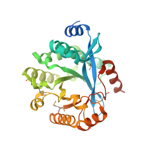
    Cyclic diguanylate (or bis-(3'-5') cyclic dimeric guanosine monophosphate; c-di-GMP) is a ubiquitous second messenger that regulates diverse cellular functions, including motility, biofilm formation, cell cycle progression, and virulence in bacteria. In the cell, degradation of c-di-GMP is catalyzed by highly specific EAL domain phosphodiesterases whose catalytic mechanism is still unclear. Here, we purified 13 EAL domain proteins from various organisms and demonstrated that their catalytic activity is associated with the presence of 10 conserved EAL domain residues. The crystal structure of the TBD1265 EAL domain was determined in free state (1.8 Å) and in complex with c-di-GMP (2.35 A), and unveiled the role of conserved residues in substrate binding and catalysis. The structure revealed the presence of two metal ions directly coordinated by six conserved residues, two oxygens of c-di-GMP phosphate, and potential catalytic water molecule. Our results support a two-metal-ion catalytic mechanism of c-di-GMP hydrolysis by EAL domain phosphodiesterases.


  • Organizational Affiliation

    Banting and Best Department of Medical Research, University of Toronto, Toronto, Ontario, Canada.


Macromolecules
Find similar proteins by:  (by identity cutoff)  |  3D Structure
Entity ID: 1
MoleculeChains Sequence LengthOrganismDetailsImage
Putative diguanylate cyclase/phosphodiesterase (GGDEF & EAL domains)
A, B
294Thiobacillus denitrificans ATCC 25259Mutation(s): 0 
Gene Names: Tbd_1265
UniProt
Find proteins for Q3SJE6 (Thiobacillus denitrificans (strain ATCC 25259))
Explore Q3SJE6 
Go to UniProtKB:  Q3SJE6
Entity Groups  
Sequence Clusters30% Identity50% Identity70% Identity90% Identity95% Identity100% Identity
UniProt GroupQ3SJE6
Sequence Annotations
Expand
  • Reference Sequence
Experimental Data & Validation

Experimental Data

  • Method: X-RAY DIFFRACTION
  • Resolution: 1.80 Å
  • R-Value Free: 0.218 
  • R-Value Work: 0.188 
  • R-Value Observed: 0.190 
  • Space Group: P 21 21 21
Unit Cell:
Length ( Å )Angle ( ˚ )
a = 50.051α = 90
b = 63.141β = 90
c = 173.282γ = 90
Software Package:
Software NamePurpose
REFMACrefinement
SBC-Collectdata collection
HKL-3000data reduction
HKL-3000data scaling
HKL-3000phasing

Structure Validation

View Full Validation Report


Entry History 

Deposition Data

Revision History  (Full details and data files)

  • Version 1.0: 2007-09-18
    Type: Initial release
  • Version 1.1: 2011-07-13
    Changes: Advisory, Source and taxonomy, Version format compliance
  • Version 1.2: 2012-10-24
    Changes: Database references