2R3W

I84V HIV-1 protease in complex with a amino decorated pyrrolidine-based inhibitor


Experimental Data Snapshot

  • Method: X-RAY DIFFRACTION
  • Resolution: 1.92 Å
  • R-Value Free: 0.231 
  • R-Value Work: 0.178 
  • R-Value Observed: 0.180 

wwPDB Validation   3D Report Full Report


Ligand Structure Quality Assessment 


This is version 1.3 of the entry. See complete history


Literature

Structural and Kinetic Analysis of Pyrrolidine-Based Inhibitors of the Drug-Resistant Ile84Val Mutant of HIV-1 Protease

Bottcher, J.Blum, A.Heine, A.Diederich, W.E.Klebe, G.

(2008) J Mol Biol 383: 347-357

  • DOI: https://doi.org/10.1016/j.jmb.2008.07.062
  • Primary Citation of Related Structures:  
    2R38, 2R3T, 2R3W, 2R43

  • PubMed Abstract: 

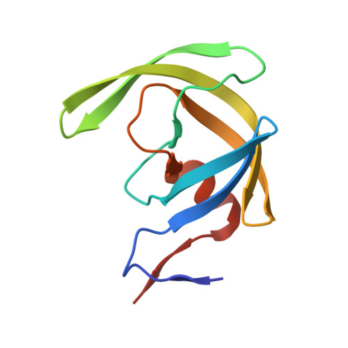
    Human immunodeficiency virus (HIV) protease is a well-established drug target in HIV chemotherapy. However, continuously increasing resistance towards approved drugs inevitably requires the development of new inhibitors preferably showing no susceptibility against resistant HIV protease strains. Recently, symmetric pyrrolidine-3,4-bis-N-benzyl-sulfonamides have been developed as a new class of HIV-1 protease inhibitors. The most promising candidate exhibited a K(i) of 74 nM towards a wild-type protease. Herein, we report the influence of the active-site mutations Ile50Val and Ile84Val on these inhibitors by structural and kinetic analysis. Although the Ile50Val mutation leads to a significant decrease in affinity for all compounds in this series, they retain or even show increased affinity towards the important Ile84Val mutation. By detailed analysis of the crystal structures of two representatives in complex with wild-type and mutant proteases, we were able to elucidate the structural basis of this phenomenon.


  • Organizational Affiliation

    Institut für Pharmazeutische Chemie, Philipps-Universität Marburg, Marbacher Weg 6, 35032 Marburg, Germany.


Macromolecules
Find similar proteins by:  (by identity cutoff)  |  3D Structure
Entity ID: 1
MoleculeChains Sequence LengthOrganismDetailsImage
Protease
A, B
99Human immunodeficiency virus 1Mutation(s): 1 
Gene Names: POL
EC: 3.4.23.16
UniProt
Find proteins for P04587 (Human immunodeficiency virus type 1 group M subtype B (isolate BH5))
Explore P04587 
Go to UniProtKB:  P04587
Entity Groups  
Sequence Clusters30% Identity50% Identity70% Identity90% Identity95% Identity100% Identity
UniProt GroupP04587
Sequence Annotations
Expand
  • Reference Sequence
Small Molecules
Ligands 2 Unique
IDChains Name / Formula / InChI Key2D Diagram3D Interactions
G3G
Query on G3G

Download Ideal Coordinates CCD File 
C [auth A]N,N'-(3S,4S)-PYRROLIDINE-3,4-DIYLBIS(4-AMINO-N-BENZYLBENZENESULFONAMIDE)
C30 H33 N5 O4 S2
XDDOZTWMJWWMBF-KYJUHHDHSA-N
CL
Query on CL

Download Ideal Coordinates CCD File 
D [auth B]CHLORIDE ION
Cl
VEXZGXHMUGYJMC-UHFFFAOYSA-M
Binding Affinity Annotations 
IDSourceBinding Affinity
G3G Binding MOAD:  2R3W Ki: 128 (nM) from 1 assay(s)
PDBBind:  2R3W Ki: 128 (nM) from 1 assay(s)
Experimental Data & Validation

Experimental Data

  • Method: X-RAY DIFFRACTION
  • Resolution: 1.92 Å
  • R-Value Free: 0.231 
  • R-Value Work: 0.178 
  • R-Value Observed: 0.180 
  • Space Group: P 21 21 2
Unit Cell:
Length ( Å )Angle ( ˚ )
a = 57.336α = 90
b = 85.763β = 90
c = 46.56γ = 90
Software Package:
Software NamePurpose
SHELXmodel building
SHELXL-97refinement
CrystalCleardata collection
HKL-2000data reduction
HKL-2000data scaling
PHASERphasing

Structure Validation

View Full Validation Report


Ligand Structure Quality Assessment 


Entry History 

Deposition Data

Revision History  (Full details and data files)

  • Version 1.0: 2008-09-02
    Type: Initial release
  • Version 1.1: 2011-07-13
    Changes: Version format compliance
  • Version 1.2: 2021-10-20
    Changes: Database references, Derived calculations
  • Version 1.3: 2023-08-30
    Changes: Data collection, Refinement description