2R0P

K252c-soaked RebC


Experimental Data Snapshot

  • Method: X-RAY DIFFRACTION
  • Resolution: 2.10 Å
  • R-Value Free: 0.255 
  • R-Value Work: 0.219 

wwPDB Validation   3D Report Full Report


Ligand Structure Quality Assessment 


This is version 1.2 of the entry. See complete history


Literature

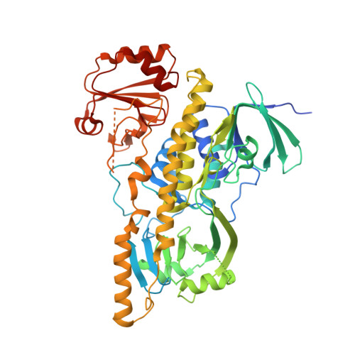
Crystallographic trapping in the rebeccamycin biosynthetic enzyme RebC

Ryan, K.S.Howard-Jones, A.R.Hamill, M.J.Elliott, S.J.Walsh, C.T.Drennan, C.L.

(2007) Proc Natl Acad Sci U S A 104: 15311-15316

  • DOI: https://doi.org/10.1073/pnas.0707190104
  • Primary Citation of Related Structures:  
    2R0C, 2R0G, 2R0P

  • PubMed Abstract: 

    The biosynthesis of rebeccamycin, an antitumor compound, involves the remarkable eight-electron oxidation of chlorinated chromopyrrolic acid. Although one rebeccamycin biosynthetic enzyme is capable of generating low levels of the eight-electron oxidation product on its own, a second protein, RebC, is required to accelerate product formation and eliminate side reactions. However, the mode of action of RebC was largely unknown. Using crystallography, we have determined a likely function for RebC as a flavin hydroxylase, captured two snapshots of its dynamic catalytic cycle, and trapped a reactive molecule, a putative substrate, in its binding pocket. These studies strongly suggest that the role of RebC is to sequester a reactive intermediate produced by its partner protein and to react with it enzymatically, preventing its conversion to a suite of degradation products that includes, at low levels, the desired product.


  • Organizational Affiliation

    Department of Biology, Massachusetts Institute of Technology, 77 Massachusetts Avenue, Cambridge, MA 02139, USA.


Macromolecules
Find similar proteins by:  (by identity cutoff)  |  3D Structure
Entity ID: 1
MoleculeChains Sequence LengthOrganismDetailsImage
RebC549Lentzea aerocolonigenesMutation(s): 0 
Gene Names: rbmDrebC
UniProt
Find proteins for Q8KI25 (Lentzea aerocolonigenes)
Explore Q8KI25 
Go to UniProtKB:  Q8KI25
Entity Groups  
Sequence Clusters30% Identity50% Identity70% Identity90% Identity95% Identity100% Identity
UniProt GroupQ8KI25
Sequence Annotations
Expand
  • Reference Sequence
Small Molecules
Ligands 3 Unique
IDChains Name / Formula / InChI Key2D Diagram3D Interactions
FAD
Query on FAD

Download Ideal Coordinates CCD File 
C [auth A]FLAVIN-ADENINE DINUCLEOTIDE
C27 H33 N9 O15 P2
VWWQXMAJTJZDQX-UYBVJOGSSA-N
K2C
Query on K2C

Download Ideal Coordinates CCD File 
D [auth A]6,7,12,13-tetrahydro-5H-indolo[2,3-a]pyrrolo[3,4-c]carbazol-5-one
C20 H13 N3 O
MEXUTNIFSHFQRG-UHFFFAOYSA-N
CL
Query on CL

Download Ideal Coordinates CCD File 
B [auth A]CHLORIDE ION
Cl
VEXZGXHMUGYJMC-UHFFFAOYSA-M
Experimental Data & Validation

Experimental Data

  • Method: X-RAY DIFFRACTION
  • Resolution: 2.10 Å
  • R-Value Free: 0.255 
  • R-Value Work: 0.219 
  • Space Group: P 1 21 1
Unit Cell:
Length ( Å )Angle ( ˚ )
a = 63.094α = 90
b = 77.509β = 108.85
c = 64.652γ = 90
Software Package:
Software NamePurpose
ADSCdata collection
CNSrefinement
DENZOdata reduction
SCALEPACKdata scaling
CNSphasing

Structure Validation

View Full Validation Report


Ligand Structure Quality Assessment 


Entry History 

Deposition Data

Revision History  (Full details and data files)

  • Version 1.0: 2007-09-25
    Type: Initial release
  • Version 1.1: 2011-07-13
    Changes: Version format compliance
  • Version 1.2: 2023-08-30
    Changes: Data collection, Database references, Derived calculations, Refinement description