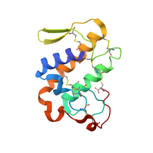
Structural Characterization of Myotoxic Ecarpholin S from Echis carinatus Venom
Zhou, X., Tan, T.C., Valiyaveettil, S., Go, M.L., Kini, R.M., Velazquez-Campoy, A., Sivaraman, J.(2008) Biophys J 95: 3366-3380
- PubMed: 18586854
- DOI: https://doi.org/10.1529/biophysj.107.117747
- Primary Citation of Related Structures:
2QHD, 2QHE, 3BJW - PubMed Abstract:
Phospholipase A(2) (PLA(2)), a common toxic component of snake venom, has been implicated in various pharmacological effects. Ecarpholin S, isolated from the venom of the snake Echis carinatus sochureki, is a phospholipase A(2) (PLA(2)) belonging to the Ser(49)-PLA(2) subgroup. It has been characterized as having low enzymatic but potent myotoxic activities. The crystal structures of native ecarpholin S and its complexes with lauric acid, and its inhibitor suramin, were elucidated. This is the first report of the structure of a member of the Ser(49)-PLA(2) subgroup. We also examined interactions of ecarpholin S with phosphatidylglycerol and lauric acid, using surface plasmon resonance, and of suramin with isothermal titration calorimetry. Most Ca(2+)-dependent PLA(2) enzymes have Asp in position 49, which plays a crucial role in Ca(2+) binding. The three-dimensional structure of ecarpholin S reveals a unique conformation of the Ca(2+)-binding loop that is not favorable for Ca(2+) coordination. Furthermore, the endogenously bound fatty acid (lauric acid) in the hydrophobic channel may also interrupt the catalytic cycle. These two observations may account for the low enzymatic activity of ecarpholin S, despite full retention of the catalytic machinery. These observations may also be applicable to other non-Asp(49)-PLA(2) enzymes. The interaction of suramin in its complex with ecarpholin S is quite different from that reported for the Lys(49)-PLA(2)/suramin complex(,) where the interfacial recognition face (i-face), C-terminal region, and N-terminal region of ecarpholin S play important roles. This study provides significant structural and functional insights into the myotoxic activity of ecarpholin S and, in general, of non-Asp(49)-PLA(2) enzymes.
Organizational Affiliation:
Department of Biological Sciences, Faculty of Science, National University of Singapore, Singapore 117543, Singapore.