2Q9T

High-resolution structure of the DING protein from Pseudomonas fluorescens


Experimental Data Snapshot

  • Method: X-RAY DIFFRACTION
  • Resolution: 1.43 Å
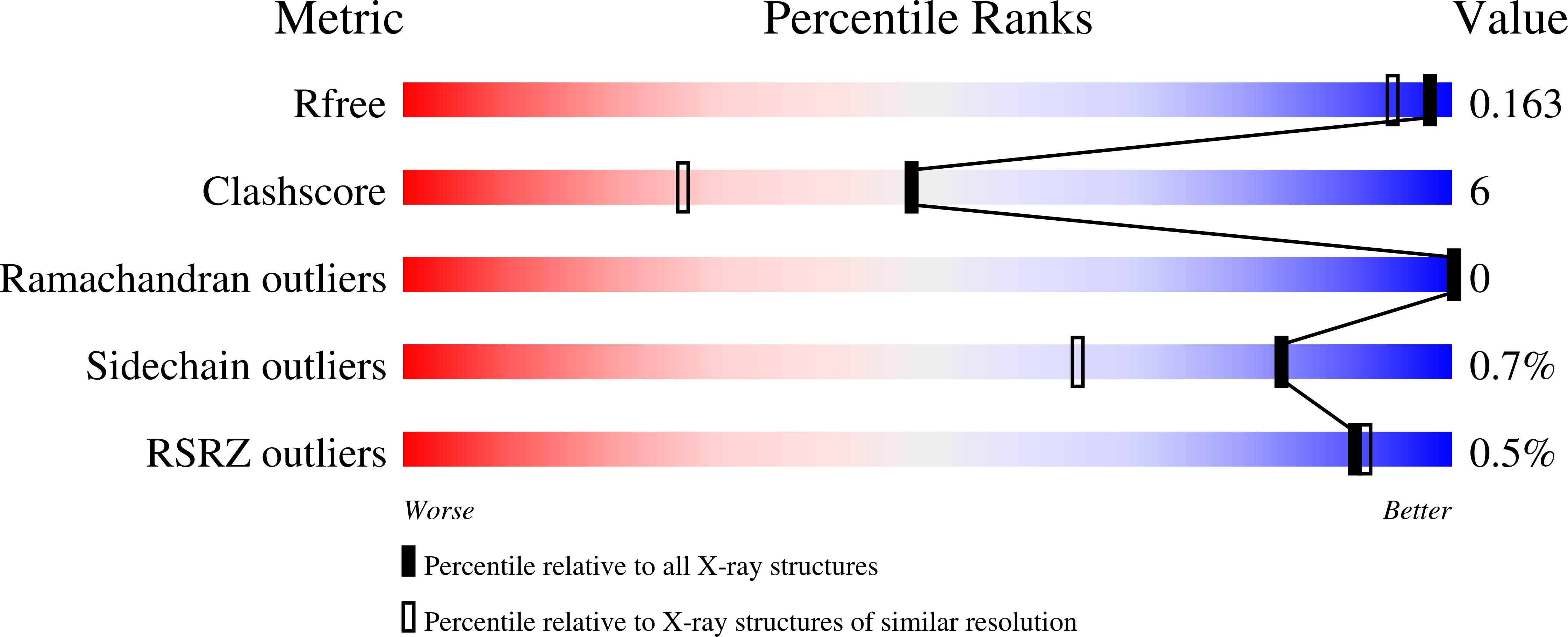
  • R-Value Free: 0.164 
  • R-Value Work: 0.131 
  • R-Value Observed: 0.133 

wwPDB Validation   3D Report Full Report


This is version 1.2 of the entry. See complete history


Literature

Structure-function relationships in a bacterial DING protein.

Ahn, S.Moniot, S.Elias, M.Chabriere, E.Kim, D.Scott, K.

(2007) FEBS Lett 581: 3455-3460

  • DOI: https://doi.org/10.1016/j.febslet.2007.06.050
  • Primary Citation of Related Structures:  
    2Q9T

  • PubMed Abstract: 

    A recombinant DING protein from Pseudomonas fluorescens has been previously shown to have a phosphate-binding site, and to be mitogenic for human cells. Here we report the three-dimensional structure of the protein, confirming a close similarity to the "Venus flytrap" structure seen in other human and bacterial phosphate-binding proteins. Site-directed mutagenesis confirms the role of a key residue involved in phosphate binding, and that the mitogenic activity is not dependent on this property. Deletion of one of the two hinged domains that constitute the Venus flytrap also eliminates phosphate binding whilst enhancing mitogenic activity.


  • Organizational Affiliation

    School of Biological Sciences, The University of Auckland, Auckland, New Zealand.


Macromolecules
Find similar proteins by:  (by identity cutoff)  |  3D Structure
Entity ID: 1
MoleculeChains Sequence LengthOrganismDetailsImage
DING379Pseudomonas fluorescensMutation(s): 0 
UniProt
Find proteins for D0VWY2 (Pseudomonas fluorescens (strain SBW25))
Explore D0VWY2 
Go to UniProtKB:  D0VWY2
Entity Groups  
Sequence Clusters30% Identity50% Identity70% Identity90% Identity95% Identity100% Identity
UniProt GroupD0VWY2
Sequence Annotations
Expand
  • Reference Sequence
Small Molecules
Ligands 5 Unique
IDChains Name / Formula / InChI Key2D Diagram3D Interactions
SO4
Query on SO4

Download Ideal Coordinates CCD File 
E [auth A],
F [auth A]
SULFATE ION
O4 S
QAOWNCQODCNURD-UHFFFAOYSA-L
PO4
Query on PO4

Download Ideal Coordinates CCD File 
B [auth A]PHOSPHATE ION
O4 P
NBIIXXVUZAFLBC-UHFFFAOYSA-K
GOL
Query on GOL

Download Ideal Coordinates CCD File 
L [auth A]GLYCEROL
C3 H8 O3
PEDCQBHIVMGVHV-UHFFFAOYSA-N
EDO
Query on EDO

Download Ideal Coordinates CCD File 
G [auth A],
H [auth A],
I [auth A],
J [auth A],
K [auth A]
1,2-ETHANEDIOL
C2 H6 O2
LYCAIKOWRPUZTN-UHFFFAOYSA-N
ACT
Query on ACT

Download Ideal Coordinates CCD File 
C [auth A],
D [auth A]
ACETATE ION
C2 H3 O2
QTBSBXVTEAMEQO-UHFFFAOYSA-M
Experimental Data & Validation

Experimental Data

  • Method: X-RAY DIFFRACTION
  • Resolution: 1.43 Å
  • R-Value Free: 0.164 
  • R-Value Work: 0.131 
  • R-Value Observed: 0.133 
  • Space Group: P 1 21 1
Unit Cell:
Length ( Å )Angle ( ˚ )
a = 36.728α = 90
b = 123.679β = 116.71
c = 40.833γ = 90
Software Package:
Software NamePurpose
REFMACrefinement
PDB_EXTRACTdata extraction
MxCuBEdata collection
XDSdata reduction
XSCALEdata scaling
MOLREPphasing

Structure Validation

View Full Validation Report


Entry History 

Deposition Data

Revision History  (Full details and data files)

  • Version 1.0: 2008-06-17
    Type: Initial release
  • Version 1.1: 2011-07-13
    Changes: Non-polymer description, Version format compliance
  • Version 1.2: 2023-08-30
    Changes: Data collection, Database references, Derived calculations, Refinement description