2Q63

HIV-1 PR mutant in complex with nelfinavir


Experimental Data Snapshot

  • Method: X-RAY DIFFRACTION
  • Resolution: 2.20 Å
  • R-Value Free: 0.250 
  • R-Value Work: 0.184 
  • R-Value Observed: 0.191 

wwPDB Validation   3D Report Full Report


Ligand Structure Quality Assessment 


This is version 1.3 of the entry. See complete history


Literature

Molecular analysis of the HIV-1 resistance development: enzymatic activities, crystal structures, and thermodynamics of nelfinavir-resistant HIV protease mutants

Kozisek, M.Bray, J.Rezacova, P.Saskova, K.Brynda, J.Pokorna, J.Mammano, F.Rulisek, L.Konvalinka, J.

(2007) J Mol Biol 374: 1005-1016

  • DOI: https://doi.org/10.1016/j.jmb.2007.09.083
  • Primary Citation of Related Structures:  
    2PYM, 2PYN, 2Q63, 2Q64

  • PubMed Abstract: 

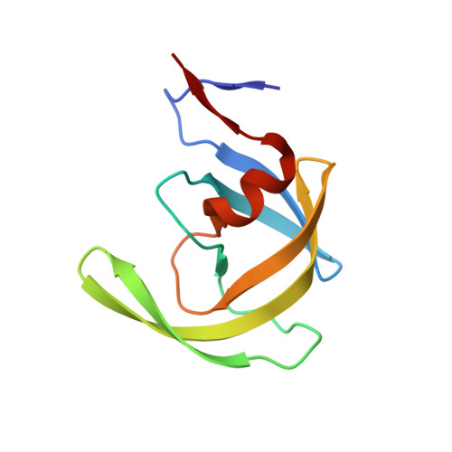
    Human immunodeficiency virus (HIV) encodes an aspartic protease (PR) that cleaves viral polyproteins into mature proteins, thus leading to the formation of infectious particles. Protease inhibitors (PIs) are successful virostatics. However, their efficiency is compromised by antiviral resistance. In the PR sequence of viral variants resistant to the PI nelfinavir, the mutations D30N and L90M appear frequently. However, these two mutations are seldom found together in vivo, suggesting that there are two alternative evolutionary pathways leading to nelfinavir resistance. Here we analyze the proteolytic activities, X-ray structures, and thermodynamics of inhibitor binding to HIV-1 PRs harboring the D30N and L90M mutations alone and in combination with other compensatory mutations. Vitality values obtained for recombinant mutant proteases and selected PR inhibitors confirm the crucial role of mutations in positions 30 and 90 for nelfinavir resistance. The combination of the D30N and L90M mutations significantly increases the enzyme vitality in the presence of nelfinavir, without a dramatic decrease in the catalytic efficiency of the recombinant enzyme. Crystal structures, molecular dynamics simulations, and calorimetric data for four mutants (D30N, D30N/A71V, D30N/N88D, and D30N/L90M) were used to augment our kinetic data. Calorimetric analysis revealed that the entropic contribution to the mutant PR/nelfinavir interaction is less favorable than the entropic contribution to the binding of nelfinavir by wild-type PR. This finding is supported by the structural data and simulations; nelfinavir binds most strongly to the wild-type protease, which has the lowest number of protein-ligand hydrogen bonds and whose structure exhibits the greatest degree of fluctuation upon inhibitor binding.


  • Organizational Affiliation

    Gilead Sciences and IOCB Research Center, Institute of Organic Chemistry and Biochemistry, Academy of Sciences of the Czech Republic, Flemingovo nám. 2, 166 10 Praha 6, Czech Republic.


Macromolecules
Find similar proteins by:  (by identity cutoff)  |  3D Structure
Entity ID: 1
MoleculeChains Sequence LengthOrganismDetailsImage
PROTEASE RETROPEPSIN
A, B
99Human immunodeficiency virus 1Mutation(s): 3 
Gene Names: gag-pol
EC: 3.4.23.16
Membrane Entity: Yes 
UniProt
Find proteins for P03367 (Human immunodeficiency virus type 1 group M subtype B (isolate BRU/LAI))
Explore P03367 
Go to UniProtKB:  P03367
Entity Groups  
Sequence Clusters30% Identity50% Identity70% Identity90% Identity95% Identity100% Identity
UniProt GroupP03367
Sequence Annotations
Expand
  • Reference Sequence
Small Molecules
Ligands 1 Unique
IDChains Name / Formula / InChI Key2D Diagram3D Interactions
1UN
Query on 1UN

Download Ideal Coordinates CCD File 
C [auth A]2-[2-HYDROXY-3-(3-HYDROXY-2-METHYL-BENZOYLAMINO)-4-PHENYL SULFANYL-BUTYL]-DECAHYDRO-ISOQUINOLINE-3-CARBOXYLIC ACID TERT-BUTYLAMIDE
C32 H45 N3 O4 S
QAGYKUNXZHXKMR-HKWSIXNMSA-N
Binding Affinity Annotations 
IDSourceBinding Affinity
1UN BindingDB:  2Q63 Ki: min: 0.07, max: 2 (nM) from 2 assay(s)
Kd: min: 3, max: 38 (nM) from 3 assay(s)
IC50: min: 1.9, max: 530 (nM) from 3 assay(s)
PDBBind:  2Q63 Kd: 66 (nM) from 1 assay(s)
Binding MOAD:  2Q63 Kd: 66 (nM) from 1 assay(s)
Experimental Data & Validation

Experimental Data

  • Method: X-RAY DIFFRACTION
  • Resolution: 2.20 Å
  • R-Value Free: 0.250 
  • R-Value Work: 0.184 
  • R-Value Observed: 0.191 
  • Space Group: P 61
Unit Cell:
Length ( Å )Angle ( ˚ )
a = 62.5α = 90
b = 62.5β = 90
c = 82.06γ = 120
Software Package:
Software NamePurpose
HKL-3000data collection
MOLREPphasing
REFMACrefinement
HKL-3000data reduction
HKL-3000data scaling

Structure Validation

View Full Validation Report


Ligand Structure Quality Assessment 


Entry History 

Deposition Data

Revision History  (Full details and data files)

  • Version 1.0: 2008-02-26
    Type: Initial release
  • Version 1.1: 2011-07-13
    Changes: Advisory, Derived calculations, Version format compliance
  • Version 1.2: 2021-10-20
    Changes: Database references, Derived calculations
  • Version 1.3: 2023-08-30
    Changes: Data collection, Refinement description