2PK5

Crystal Structure of HIV-1 Protease (Q7K, L33I, L63I ) in Complex with KNI-10075


Experimental Data Snapshot

  • Method: X-RAY DIFFRACTION
  • Resolution: 1.90 Å
  • R-Value Free: 0.200 
  • R-Value Work: 0.160 
  • R-Value Observed: 0.164 

wwPDB Validation   3D Report Full Report


This is version 1.6 of the entry. See complete history


Literature

Compensating enthalpic and entropic changes hinder binding affinity optimization.

Lafont, V.Armstrong, A.A.Ohtaka, H.Kiso, Y.Mario Amzel, L.Freire, E.

(2007) Chem Biol Drug Des 69: 413-422

  • DOI: https://doi.org/10.1111/j.1747-0285.2007.00519.x
  • Primary Citation of Related Structures:  
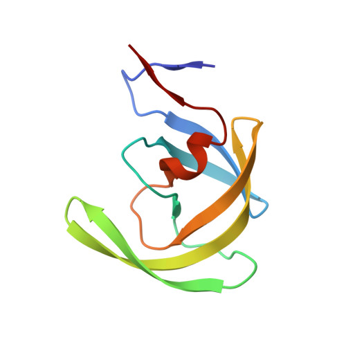
    2PK5, 2PK6

  • PubMed Abstract: 

    A common strategy to improve the potency of drug candidates is to introduce chemical functionalities, like hydrogen bond donors or acceptors, at positions where they are able to establish strong interactions with the target. However, it is often observed that the added functionalities do not necessarily improve potency even if they form strong hydrogen bonds. Here, we explore the thermodynamic and structural basis for those observations. KNI-10033 is a potent experimental HIV-1 protease inhibitor with picomolar affinity against the wild-type enzyme (K(d) = 13 pm). The potency of the inhibitor is the result of favorable enthalpic (DeltaH = -8.2 kcal/mol) and entropic (-TDeltaS = -6.7 kcal/mol) interactions. The replacement of the thioether group in KNI-10033 by a sulfonyl group (KNI-10075) results in a strong hydrogen bond with the amide of Asp 30B of the HIV-1 protease. This additional hydrogen bond improves the binding enthalpy by 3.9 kcal/mol; however, the enthalpy gain is completely compensated by an entropy loss, resulting in no affinity change. Crystallographic and thermodynamic analysis of the inhibitor/protease complexes indicates that the entropy losses are due to a combination of conformational and solvation effects. These results provide a set of practical guidelines aimed at overcoming enthalpy/entropy compensation and improve binding potency.


  • Organizational Affiliation

    Department of Biology, Johns Hopkins University, Baltimore, MD 21218, USA.


Macromolecules
Find similar proteins by:  (by identity cutoff)  |  3D Structure
Entity ID: 1
MoleculeChains Sequence LengthOrganismDetailsImage
Protease
A, B
99Human immunodeficiency virus 1Mutation(s): 3 
Gene Names: gag-pol
UniProt
Find proteins for Q9J2P7 (Human immunodeficiency virus 1)
Explore Q9J2P7 
Go to UniProtKB:  Q9J2P7
Entity Groups  
Sequence Clusters30% Identity50% Identity70% Identity90% Identity95% Identity100% Identity
UniProt GroupQ9J2P7
Sequence Annotations
Expand
  • Reference Sequence
Small Molecules
Ligands 2 Unique
IDChains Name / Formula / InChI Key2D Diagram3D Interactions
075
Query on 075

Download Ideal Coordinates CCD File 
C [auth A](4R)-N-[(1S,2R)-2-hydroxy-2,3-dihydro-1H-inden-1-yl]-3-[(2S,3S)-2-hydroxy-3-({N-[(isoquinolin-5-yloxy)acetyl]-3-(methyl sulfonyl)-L-alanyl}amino)-4-phenylbutanoyl]-5,5-dimethyl-1,3-thiazolidine-4-carboxamide
C40 H45 N5 O9 S2
GNSJNHNIJIEWNZ-SHJYOGRKSA-N
GOL
Query on GOL

Download Ideal Coordinates CCD File 
D [auth A]GLYCEROL
C3 H8 O3
PEDCQBHIVMGVHV-UHFFFAOYSA-N
Binding Affinity Annotations 
IDSourceBinding Affinity
075 Binding MOAD:  2PK5 Kd: 0.02 (nM) from 1 assay(s)
Biologically Interesting Molecules (External Reference) 1 Unique
Entity ID: 2
IDChains NameType/Class2D Diagram3D Interactions
PRD_000576 (075)
Query on PRD_000576
C [auth A]KNI-10075Peptide-like / Inhibitor
Experimental Data & Validation

Experimental Data

  • Method: X-RAY DIFFRACTION
  • Resolution: 1.90 Å
  • R-Value Free: 0.200 
  • R-Value Work: 0.160 
  • R-Value Observed: 0.164 
  • Space Group: P 21 21 2
Unit Cell:
Length ( Å )Angle ( ˚ )
a = 58.842α = 90
b = 85.991β = 90
c = 46.21γ = 90
Software Package:
Software NamePurpose
DENZOdata reduction
SCALEPACKdata scaling
REFMACrefinement
PDB_EXTRACTdata extraction
HKL-2000data reduction
MOLREPphasing

Structure Validation

View Full Validation Report


Entry History 

Deposition Data

Revision History  (Full details and data files)

  • Version 1.0: 2007-05-08
    Type: Initial release
  • Version 1.1: 2008-05-01
    Changes: Version format compliance
  • Version 1.2: 2011-07-13
    Changes: Atomic model, Database references, Derived calculations, Non-polymer description, Structure summary, Version format compliance
  • Version 1.3: 2012-12-12
    Changes: Other
  • Version 1.4: 2017-10-18
    Changes: Refinement description
  • Version 1.5: 2021-10-20
    Changes: Database references, Derived calculations, Structure summary
  • Version 1.6: 2023-08-30
    Changes: Data collection, Refinement description