2OYP

T Cell Immunoglobulin Mucin-3 Crystal Structure Revealed a Galectin-9-independent Binding Surface


Experimental Data Snapshot

  • Method: X-RAY DIFFRACTION
  • Resolution: 1.95 Å
  • R-Value Free: 0.200 
  • R-Value Work: 0.156 
  • R-Value Observed: 0.158 

wwPDB Validation   3D Report Full Report


This is version 1.5 of the entry. See complete history


Literature

T cell immunoglobulin mucin-3 crystal structure reveals a galectin-9-independent ligand-binding surface

Cao, E.Zang, X.Ramagopal, U.A.Mukhopadhaya, A.Fedorov, A.A.Fedorov, E.V.Zencheck, W.D.Lary, J.W.Cole, J.L.Deng, H.Xiao, H.Dilorenzo, T.P.Allison, J.P.Nathenson, S.G.Almo, S.C.

(2007) Immunity 26: 311-321

  • DOI: https://doi.org/10.1016/j.immuni.2007.01.016
  • Primary Citation of Related Structures:  
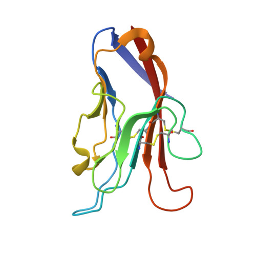
    2OYP

  • PubMed Abstract: 

    The T cell immunoglobulin mucin (Tim) family of receptors regulates effector CD4(+) T cell functions and is implicated in autoimmune and allergic diseases. Tim-3 induces immunological tolerance, and engagement of the Tim-3 immunoglobulin variable (IgV) domain by galectin-9 is important for appropriate termination of T helper 1-immune responses. The 2 A crystal structure of the Tim-3 IgV domain demonstrated that four cysteines, which are invariant within the Tim family, form two noncanonical disulfide bonds, resulting in a surface not present in other immunoglobulin superfamily members. Biochemical and biophysical studies demonstrated that this unique structural feature mediates a previously unidentified galectin-9-independent binding process and suggested that this structural feature is conserved within the entire Tim family. The current work provided a graphic example of the relationship between sequence, structure, and function and suggested that the interplay between multiple Tim-3-binding activities contributes to the regulated assembly of signaling complexes required for effective Th1-mediated immunity.


  • Organizational Affiliation

    Department of Cell Biology, Albert Einstein College of Medicine, Bronx, NY 10461, USA.


Macromolecules
Find similar proteins by:  (by identity cutoff)  |  3D Structure
Entity ID: 1
MoleculeChains Sequence LengthOrganismDetailsImage
Hepatitis A virus cellular receptor 2109Mus musculusMutation(s): 0 
Gene Names: Havcr2
UniProt & NIH Common Fund Data Resources
Find proteins for Q3KP82 (Mus musculus)
Explore Q3KP82 
Go to UniProtKB:  Q3KP82
IMPC:  MGI:2159682
Entity Groups  
Sequence Clusters30% Identity50% Identity70% Identity90% Identity95% Identity100% Identity
UniProt GroupQ3KP82
Sequence Annotations
Expand
  • Reference Sequence
Small Molecules
Ligands 1 Unique
IDChains Name / Formula / InChI Key2D Diagram3D Interactions
SO4
Query on SO4

Download Ideal Coordinates CCD File 
B [auth A]SULFATE ION
O4 S
QAOWNCQODCNURD-UHFFFAOYSA-L
Experimental Data & Validation

Experimental Data

  • Method: X-RAY DIFFRACTION
  • Resolution: 1.95 Å
  • R-Value Free: 0.200 
  • R-Value Work: 0.156 
  • R-Value Observed: 0.158 
  • Space Group: P 1 21 1
Unit Cell:
Length ( Å )Angle ( ˚ )
a = 23α = 90
b = 52.809β = 100.94
c = 38.941γ = 90
Software Package:
Software NamePurpose
DENZOdata reduction
SCALEPACKdata scaling
REFMACrefinement
PDB_EXTRACTdata extraction
CBASSdata collection
HKL-2000data reduction
MOLREPphasing

Structure Validation

View Full Validation Report


Entry History 

Deposition Data

Revision History  (Full details and data files)

  • Version 1.0: 2007-04-10
    Type: Initial release
  • Version 1.1: 2008-05-01
    Changes: Version format compliance
  • Version 1.2: 2011-07-13
    Changes: Version format compliance
  • Version 1.3: 2013-11-13
    Changes: Database references
  • Version 1.4: 2017-10-18
    Changes: Refinement description
  • Version 1.5: 2023-08-30
    Changes: Data collection, Database references, Derived calculations, Refinement description