2OX9

Mouse Scavenger Receptor C-type Lectin carbohydrate-recognition domain.


Experimental Data Snapshot

  • Method: X-RAY DIFFRACTION
  • Resolution: 1.95 Å
  • R-Value Free: 0.273 
  • R-Value Work: 0.224 

wwPDB Validation   3D Report Full Report


This is version 2.0 of the entry. See complete history


Literature

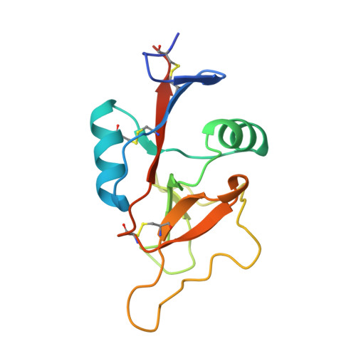
Scavenger receptor C-type lectin binds to the leukocyte cell surface glycan Lewis(x) by a novel mechanism.

Feinberg, H.Taylor, M.E.Weis, W.I.

(2007) J Biol Chem 282: 17250-17258

  • DOI: https://doi.org/10.1074/jbc.M701624200
  • Primary Citation of Related Structures:  
    2OX8, 2OX9

  • PubMed Abstract: 

    The scavenger receptor C-type lectin (SRCL) is unique in the family of class A scavenger receptors, because in addition to binding sites for oxidized lipoproteins it also contains a C-type carbohydrate-recognition domain (CRD) that interacts with specific glycans. Both human and mouse SRCL are highly specific for the Lewis(x) trisaccharide, which is commonly found on the surfaces of leukocytes and some tumor cells. Structural analysis of the CRD of mouse SRCL in complex with Lewis(x) and mutagenesis show the basis for this specificity. The interaction between mouse SRCL and Lewis(x) is analogous to the way that selectins and DC-SIGN bind to related fucosylated glycans, but the mechanism of the interaction is novel, because it is based on a primary galactose-binding site similar to the binding site in the asialoglycoprotein receptor. Crystals of the human receptor lacking bound calcium ions reveal an alternative conformation in which a glycan ligand would be released during receptor-mediated endocytosis.


  • Organizational Affiliation

    Department of Structural Biology, Stanford University School of Medicine, Stanford, California 94306, USA.


Macromolecules
Find similar proteins by:  (by identity cutoff)  |  3D Structure
Entity ID: 1
MoleculeChains Sequence LengthOrganismDetailsImage
Collectin placenta 1
A, B, C, D
140Mus musculusMutation(s): 0 
Gene Names: Colec12CL-P1
UniProt & NIH Common Fund Data Resources
Find proteins for Q8K4Q8 (Mus musculus)
Explore Q8K4Q8 
Go to UniProtKB:  Q8K4Q8
IMPC:  MGI:2152907
Entity Groups  
Sequence Clusters30% Identity50% Identity70% Identity90% Identity95% Identity100% Identity
UniProt GroupQ8K4Q8
Sequence Annotations
Expand
  • Reference Sequence
Oligosaccharides

Help

Entity ID: 2
MoleculeChains Length2D Diagram Glycosylation3D Interactions
alpha-L-fucopyranose-(1-3)-[beta-D-galactopyranose-(1-4)]2-acetamido-2-deoxy-beta-D-glucopyranose
E, F, G, H
3N/A
Glycosylation Resources
GlyTouCan:  G00051MO
GlyCosmos:  G00051MO
GlyGen:  G00051MO
Small Molecules
Ligands 1 Unique
IDChains Name / Formula / InChI Key2D Diagram3D Interactions
CA
Query on CA

Download Ideal Coordinates CCD File 
I [auth A]
J [auth A]
K [auth A]
L [auth A]
M [auth B]
I [auth A],
J [auth A],
K [auth A],
L [auth A],
M [auth B],
N [auth B],
O [auth B],
P [auth B],
Q [auth C],
R [auth C],
S [auth C],
T [auth C],
U [auth D],
V [auth D],
W [auth D],
X [auth D]
CALCIUM ION
Ca
BHPQYMZQTOCNFJ-UHFFFAOYSA-N
Experimental Data & Validation

Experimental Data

  • Method: X-RAY DIFFRACTION
  • Resolution: 1.95 Å
  • R-Value Free: 0.273 
  • R-Value Work: 0.224 
  • Space Group: P 1
Unit Cell:
Length ( Å )Angle ( ˚ )
a = 47.998α = 67.75
b = 53.755β = 76.7
c = 59.084γ = 85.37
Software Package:
Software NamePurpose
CNSrefinement
Blu-Icedata collection
COMOphasing

Structure Validation

View Full Validation Report


Entry History 

Deposition Data

Revision History  (Full details and data files)

  • Version 1.0: 2007-04-03
    Type: Initial release
  • Version 1.1: 2008-05-01
    Changes: Version format compliance
  • Version 1.2: 2011-07-13
    Changes: Non-polymer description, Version format compliance
  • Version 2.0: 2020-07-29
    Type: Remediation
    Reason: Carbohydrate remediation
    Changes: Atomic model, Data collection, Derived calculations, Structure summary