2NYN

Crystal structure of phenylalanine ammonia-lyase from Anabaena variabilis


Experimental Data Snapshot

  • Method: X-RAY DIFFRACTION
  • Resolution: 1.90 Å
  • R-Value Free: 0.242 
  • R-Value Work: 0.223 

wwPDB Validation   3D Report Full Report


This is version 2.0 of the entry. See complete history


Literature

Discovery of Two Cyanobacterial Phenylalanine Ammonia Lyases: Kinetic and Structural Characterization.

Moffitt, M.C.Louie, G.V.Bowman, M.E.Pence, J.Noel, J.P.Moore, B.S.

(2007) Biochemistry 46: 1004-1012

  • DOI: https://doi.org/10.1021/bi061774g
  • Primary Citation of Related Structures:  
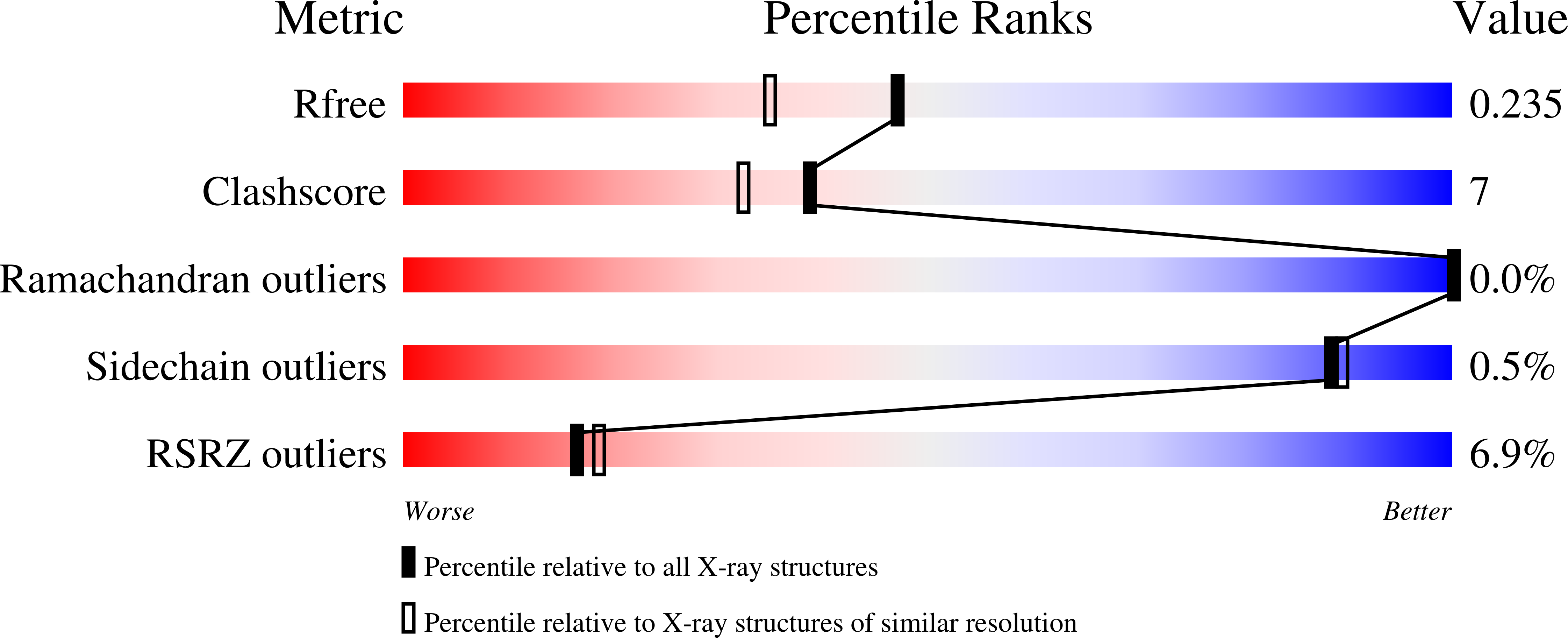
    2NYF, 2NYN

  • PubMed Abstract: 

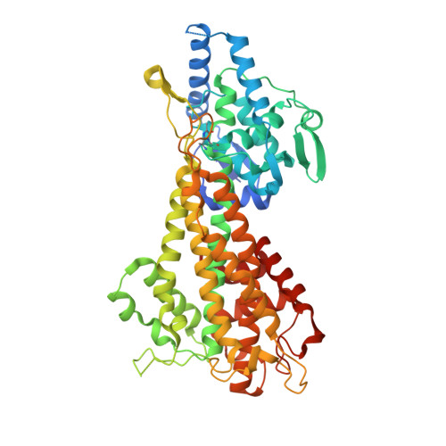
    Phenylalanine ammonia lyase (PAL) catalyzes the deamination of phenylalanine to cinnamate and ammonia. While PALs are common in terrestrial plants where they catalyze the first committed step in the formation of phenylpropanoids, only a few prokaryotic PALs have been identified to date. Here we describe for the first time PALs from cyanobacteria, in particular, Anabaena variabilis ATCC 29413 and Nostoc punctiforme ATCC 29133, identified by screening the genome sequences of these organisms for members of the aromatic amino acid ammonia lyase family. Both PAL genes associate with secondary metabolite biosynthetic gene clusters as observed for other eubacterial PAL genes. In comparison to eukaryotic homologues, the cyanobacterial PALs are 20% smaller in size but share similar substrate selectivity and kinetic activity toward L-phenylalanine over L-tyrosine. Structure elucidation by protein X-ray crystallography confirmed that the two cyanobacterial PALs are similar in tertiary and quatenary structure to plant and yeast PALs as well as the mechanistically related histidine ammonia lyases.


  • Organizational Affiliation

    Scripps Institution of Oceanography and Skaggs School of Pharmacy and Pharmaceutical Sciences, University of California at San Diego, La Jolla, California 92093, USA.


Macromolecules
Find similar proteins by:  (by identity cutoff)  |  3D Structure
Entity ID: 1
MoleculeChains Sequence LengthOrganismDetailsImage
Phenylalanine/histidine ammonia-lyase
A, B, C, D
565Trichormus variabilisMutation(s): 0 
EC: 4.3.1.3
UniProt
Find proteins for Q3M5Z3 (Trichormus variabilis (strain ATCC 29413 / PCC 7937))
Explore Q3M5Z3 
Go to UniProtKB:  Q3M5Z3
Entity Groups  
Sequence Clusters30% Identity50% Identity70% Identity90% Identity95% Identity100% Identity
UniProt GroupQ3M5Z3
Sequence Annotations
Expand
  • Reference Sequence
Small Molecules
Modified Residues  1 Unique
IDChains TypeFormula2D DiagramParent
MDO
Query on MDO
A, B, C, D
L-PEPTIDE LINKINGC8 H11 N3 O3ALA, SER, GLY
Experimental Data & Validation

Experimental Data

  • Method: X-RAY DIFFRACTION
  • Resolution: 1.90 Å
  • R-Value Free: 0.242 
  • R-Value Work: 0.223 
  • Space Group: P 1
Unit Cell:
Length ( Å )Angle ( ˚ )
a = 78.193α = 103.52
b = 88.515β = 97.82
c = 90.058γ = 116.24
Software Package:
Software NamePurpose
MOLREPphasing
CNSrefinement
XDSdata reduction
XSCALEdata scaling

Structure Validation

View Full Validation Report


Entry History 

Deposition Data

Revision History  (Full details and data files)

  • Version 1.0: 2007-02-06
    Type: Initial release
  • Version 1.1: 2008-05-01
    Changes: Version format compliance
  • Version 1.2: 2011-07-13
    Changes: Version format compliance
  • Version 1.3: 2023-08-30
    Changes: Data collection, Database references, Derived calculations, Refinement description
  • Version 2.0: 2023-11-15
    Changes: Atomic model, Data collection, Derived calculations