Structural studies on Plasmodium vivax merozoite surface protein-1
Babon, J.J., Morgan, W.D., Kelly, G., Eccleston, J.F., Feeney, J., Holder, A.A.(2007) Mol Biochem Parasitol 153: 31-40
- PubMed: 17343930
- DOI: https://doi.org/10.1016/j.molbiopara.2007.01.015
- Primary Citation of Related Structures:
2NPR - PubMed Abstract:
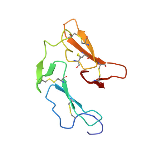
Plasmodium vivax infection is the second most common cause of malaria throughout the world. Like other Plasmodium species, P. vivax has a large protein complex, MSP-1, located on the merozoite surface. The C-terminal MSP-1 sub-unit, MSP-1(42), is cleaved during red blood cell invasion, causing the majority of the complex to be shed and leaving only a small 15kDa sub-unit, MSP-1(19), on the merozite surface. MSP-1(19) is considered a strong vaccine candidate. We have determined the solution structure of MSP-1(19) from P. vivax using nuclear magnetic resonance (NMR) and show that, like in other Plasmodium species, it consists of two EGF-like domains that are oriented head-to-tail. The protein has a flat, disk-like shape with a highly charged surface. When MSP-1(19) is part of the larger MSP-1(42) precursor it exists as an independent domain with no stable contacts to the rest of the sub-unit. Gel filtration and analytical ultracentrifugation experiments indicate that P. vivax MSP-1(42) exists as a dimer in solution. MSP-1(19) itself is a monomer, however, 35 amino-acids immediately upstream of its N-terminus are sufficient to cause dimerization. Our data suggest that if MSP-1(42) exists as a dimer in vivo, secondary processing would cause the dissociation of two tightly linked MSP-1(19) proteins on the merozoite surface just prior to invasion.
Organizational Affiliation:
Division of Parasitology, National Institute for Medical Research, The Ridgeway, Mill Hill, London NW7 1AA, United Kingdom. babon@wehi.EDU.AU