High-resolution NMR structure of the antimicrobial peptide protegrin-2 in the presence of DPC micelles.
Usachev, K.S., Efimov, S.V., Kolosova, O.A., Filippov, A.V., Klochkov, V.V.(2015) J Biomol NMR 61: 227-234
- PubMed: 25430060
- DOI: https://doi.org/10.1007/s10858-014-9885-4
- Primary Citation of Related Structures:
2MUH - PubMed Abstract:
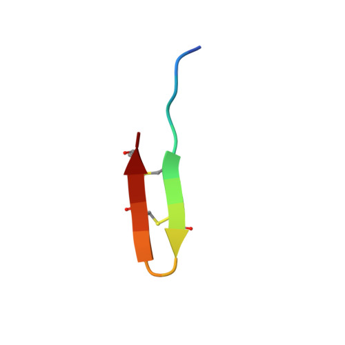
PG-1 adopts a dimeric structure in dodecylphosphocholine (DPC) micelles, and a channel is formed by the association of several dimers but the molecular mechanisms of the membrane damage by non-α-helical peptides are still unknown. The formation of the PG-1 dimer is important for pore formation in the lipid bilayer, since the dimer can be regarded as the primary unit for assembly into the ordered aggregates. It was supposed that only 12 residues (RGGRL-CYCRR-RFCVC-V) are needed to endow protegrin molecules with strong antibacterial activity and that at least four additional residues are needed to add potent antifungal properties. Thus, the 16-residue protegrin (PG-2) represents the minimal structure needed for broad-spectrum antimicrobial activity encompassing bacteria and fungi. As the peptide conformation and peptide-to-membrane binding properties are very sensitive to single amino acid substitutions, the solution structure of PG-2 in solution and in a membrane mimicking environment are crucial. In order to find evidence if the oligomerization state of PG-1 in a lipid environment will be the same or not for another protegrins, we investigate in the present work the PG-2 NMR solution structure in the presence of perdeuterated DPC micelles. The NMR study reported in the present work indicates that PG-2 form a well-defined structure (PDB: 2MUH) composed of a two-stranded antiparallel β-sheet when it binds to DPC micelles.
Organizational Affiliation:
Kazan Federal University, Kremlevskaya, 18, Kazan, 420008, Russian Federation, k.usachev@kpfu.ru.