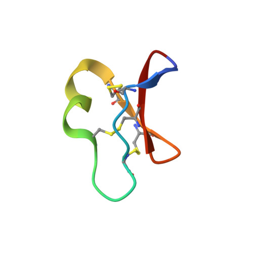
Despite a conserved cystine knot motif, different cyclotides have different membrane binding modes.
Wang, C.K., Colgrave, M.L., Ireland, D.C., Kaas, Q., Craik, D.J.(2009) Biophys J 97: 1471-1481
- PubMed: 19720036
- DOI: https://doi.org/10.1016/j.bpj.2009.06.032
- Primary Citation of Related Structures:
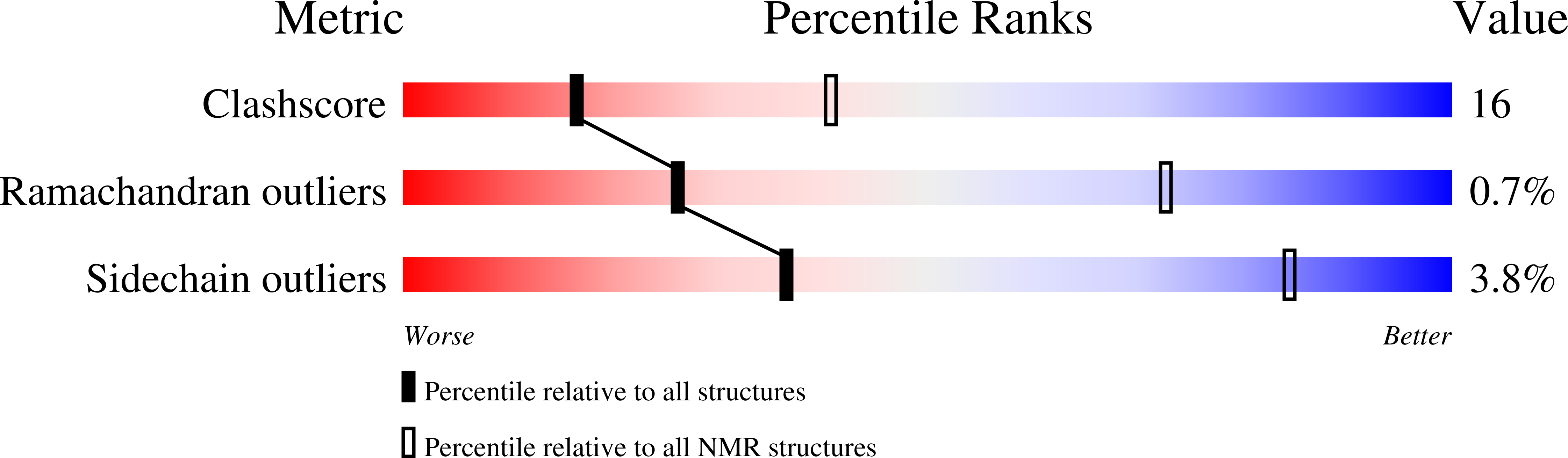
2KCG, 2KCH - PubMed Abstract:
Cyclotides are cyclic proteins produced by plants for defense against pests. Because of their remarkable stability and diverse bioactivities, they have a range of potential therapeutic applications. The bioactivities of cyclotides are believed to be mediated through membrane interactions. To determine the structural basis for the biological activity of the two major subfamilies of cyclotides, we determined the conformation and orientation of kalata B2 (kB2), a Möbius cyclotide, and cycloviolacin O2 (cO2), a bracelet cyclotide, bound to dodecylphosphocholine micelles, using NMR spectroscopy in the presence and absence of 5- and 16-doxylstearate relaxation probes. Analysis of binding curves using the Langmuir isotherm indicated that cO2 and kB2 have association constants of 7.0 x 10(3) M(-1) and 6.0 x 10(3) M(-1), respectively, consistent with the notion that they are bound near the surface, rather than buried deeply within the micelle. This suggestion is supported by the selective broadening of micelle-bound cyclotide NMR signals upon addition of paramagnetic Mn ions. The cyclotides from the different subfamilies exhibited clearly different binding orientations at the micelle surface. Structural analysis of cO2 confirmed that the main element of the secondary structure is a beta-hairpin centered in loop 5. A small helical turn is present in loop 3. Analysis of the surface profile of cO2 shows that a hydrophobic patch stretches over loops 2 and 3, in contrast to the hydrophobic patch of kB2, which predominantly involves loops 2 and 5. The different location of the hydrophobic patches in the two cyclotides explains their different binding orientations and provides an insight into the biological activities of cyclotides.
Organizational Affiliation:
University of Queensland, Institute for Molecular Bioscience, Brisbane, Queensland, Australia.