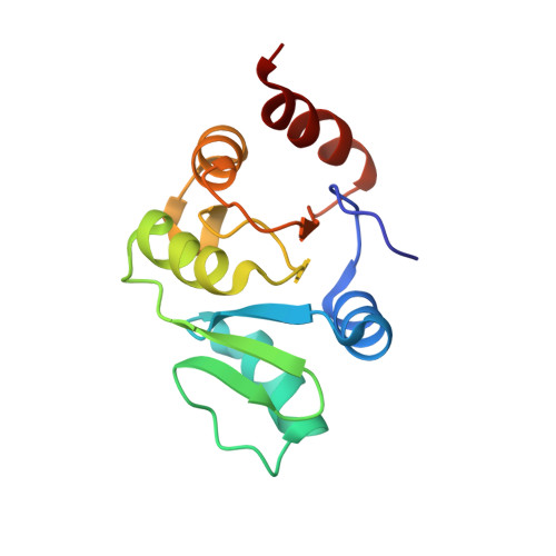
Nuclear magnetic resonance structure shows that the severe acute respiratory syndrome coronavirus-unique domain contains a macrodomain fold.
Chatterjee, A., Johnson, M.A., Serrano, P., Pedrini, B., Joseph, J.S., Neuman, B.W., Saikatendu, K., Buchmeier, M.J., Kuhn, P., Wuthrich, K.(2009) J Virol 83: 1823-1836
- PubMed: 19052085
- DOI: https://doi.org/10.1128/JVI.01781-08
- Primary Citation of Related Structures:
2JZD, 2JZE, 2JZF, 2RNK - PubMed Abstract:
The nuclear magnetic resonance (NMR) structure of a central segment of the previously annotated severe acute respiratory syndrome (SARS)-unique domain (SUD-M, for "middle of the SARS-unique domain") in SARS coronavirus (SARS-CoV) nonstructural protein 3 (nsp3) has been determined. SUD-M(513-651) exhibits a macrodomain fold containing the nsp3 residues 528 to 648, and there is a flexibly extended N-terminal tail with the residues 513 to 527 and a C-terminal flexible tail of residues 649 to 651. As a follow-up to this initial result, we also solved the structure of a construct representing only the globular domain of residues 527 to 651 [SUD-M(527-651)]. NMR chemical shift perturbation experiments showed that SUD-M(527-651) binds single-stranded poly(A) and identified the contact area with this RNA on the protein surface, and electrophoretic mobility shift assays then confirmed that SUD-M has higher affinity for purine bases than for pyrimidine bases. In a further search for clues to the function, we found that SUD-M(527-651) has the closest three-dimensional structure homology with another domain of nsp3, the ADP-ribose-1"-phosphatase nsp3b, although the two proteins share only 5% sequence identity in the homologous sequence regions. SUD-M(527-651) also shows three-dimensional structure homology with several helicases and nucleoside triphosphate-binding proteins, but it does not contain the motifs of catalytic residues found in these structural homologues. The combined results from NMR screening of potential substrates and the structure-based homology studies now form a basis for more focused investigations on the role of the SARS-unique domain in viral infection.
Organizational Affiliation:
Department of Molecular Biology, Skaggs Institute for Chemical Biology, The Scripps Research Institute, La Jolla, California 92037, USA.