2JCD

Structure of the N-oxygenase AurF from Streptomyces thioluteus


Experimental Data Snapshot

  • Method: X-RAY DIFFRACTION
  • Resolution: 2.11 Å
  • R-Value Free: 0.242 
  • R-Value Work: 0.196 
  • R-Value Observed: 0.198 

wwPDB Validation   3D Report Full Report


This is version 1.3 of the entry. See complete history


Literature

Structure and Action of the N-Oxygenase Aurf from Streptomyces Thioluteus.

Zocher, G.E.Winkler, R.Hertweck, C.Schulz, G.E.

(2007) J Mol Biol 373: 65

  • DOI: https://doi.org/10.1016/j.jmb.2007.06.014
  • Primary Citation of Related Structures:  
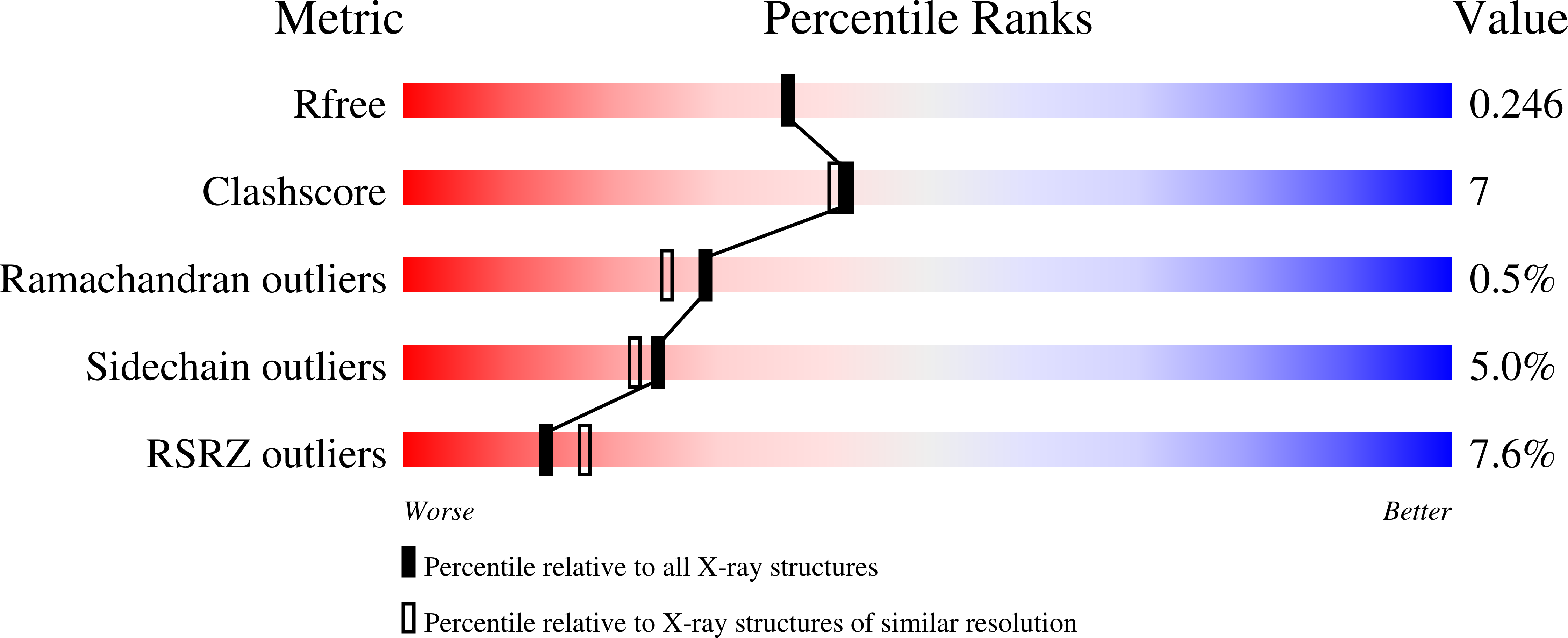
    2JCD

  • PubMed Abstract: 

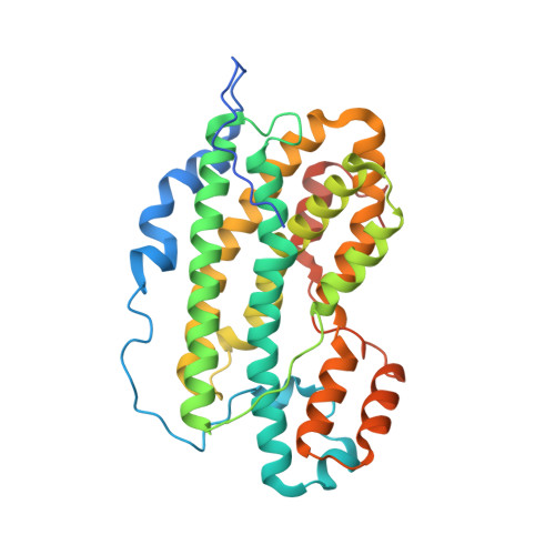
    Nitro groups are found in a number of bioactive compounds. Most of them arise by a stepwise mono-oxygenation of amino groups. One of the involved enzymes is AurF participating in the biosynthesis of aureothin. Its structure was established at 2.1 A resolution showing a homodimer with a binuclear manganese cluster. The enzyme preparation, which yielded the analyzed crystals, showed activity using in vitro and in vivo assays. Chain fold and cluster are homologous with ribonucleotide reductase subunit R2 and related enzymes. The two manganese ions and an iron content of about 15% were established by anomalous X-ray diffraction. A comparison of the cluster with more common di-iron clusters suggested an additional histidine in the coordination sphere to cause the preference for manganese over iron. There is no oxo-bridge. The substrate p-amino-benzoate was modeled into the active center. The model is supported by mutant activity measurements. It shows the geometry of the reaction and explains the established substrate spectrum.


  • Organizational Affiliation

    Institut für Organische Chemie und Biochemie, Albert-Ludwigs-Universität, Albertstrasse 21, 79104 Freiburg im Breisgau, Germany.


Macromolecules
Find similar proteins by:  (by identity cutoff)  |  3D Structure
Entity ID: 1
MoleculeChains Sequence LengthOrganismDetailsImage
N-OXIDASE
A, B
345Streptomyces thioluteusMutation(s): 0 
UniProt
Find proteins for Q70KH9 (Streptomyces thioluteus)
Explore Q70KH9 
Go to UniProtKB:  Q70KH9
Entity Groups  
Sequence Clusters30% Identity50% Identity70% Identity90% Identity95% Identity100% Identity
UniProt GroupQ70KH9
Sequence Annotations
Expand
  • Reference Sequence
Small Molecules
Ligands 2 Unique
IDChains Name / Formula / InChI Key2D Diagram3D Interactions
EDO
Query on EDO

Download Ideal Coordinates CCD File 
E [auth A]
F [auth A]
G [auth A]
H [auth A]
I [auth A]
E [auth A],
F [auth A],
G [auth A],
H [auth A],
I [auth A],
L [auth B],
M [auth B]
1,2-ETHANEDIOL
C2 H6 O2
LYCAIKOWRPUZTN-UHFFFAOYSA-N
MN
Query on MN

Download Ideal Coordinates CCD File 
C [auth A],
D [auth A],
J [auth B],
K [auth B]
MANGANESE (II) ION
Mn
WAEMQWOKJMHJLA-UHFFFAOYSA-N
Experimental Data & Validation

Experimental Data

  • Method: X-RAY DIFFRACTION
  • Resolution: 2.11 Å
  • R-Value Free: 0.242 
  • R-Value Work: 0.196 
  • R-Value Observed: 0.198 
  • Space Group: P 21 21 21
Unit Cell:
Length ( Å )Angle ( ˚ )
a = 62.222α = 90
b = 115.438β = 90
c = 125.801γ = 90
Software Package:
Software NamePurpose
ARP/wARPmodel building
XDSdata reduction
SHELXphasing
SHARPphasing
PHASERphasing
ARP/wARPphasing
REFMACphasing
Cootphasing
REFMACrefinement

Structure Validation

View Full Validation Report


Entry History 

Deposition Data

Revision History  (Full details and data files)

  • Version 1.0: 2007-09-11
    Type: Initial release
  • Version 1.1: 2011-05-08
    Changes: Version format compliance
  • Version 1.2: 2011-07-13
    Changes: Version format compliance
  • Version 1.3: 2017-06-28
    Changes: Refinement description