2J9J

Atomic-resolution Crystal Structure of Chemically-Synthesized HIV-1 Protease Complexed with Inhibitor JG-365


Experimental Data Snapshot

  • Method: X-RAY DIFFRACTION
  • Resolution: 1.04 Å
  • R-Value Free: 0.191 
  • R-Value Observed: 0.142 

wwPDB Validation   3D Report Full Report


This is version 2.1 of the entry. See complete history


Literature

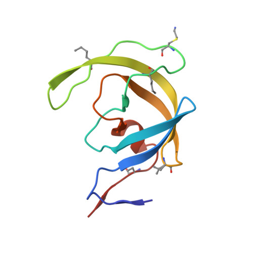
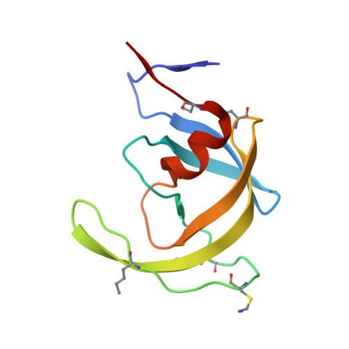
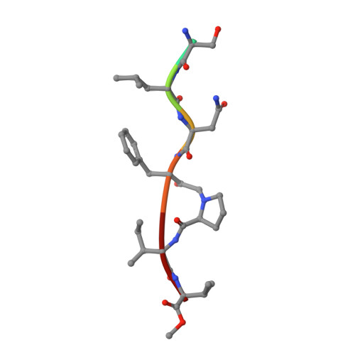
Insights from Atomic-Resolution X-Ray Structures of Chemically Synthesized HIV-1 Protease in Complex with Inhibitors.

Johnson, E.C.B.Malito, E.Shen, Y.Pentelute, B.Rich, D.Florian, J.Tang, W.J.Kent, S.B.H.

(2007) J Mol Biol 373: 573

  • DOI: https://doi.org/10.1016/j.jmb.2007.07.054
  • Primary Citation of Related Structures:  
    2J9J, 2J9K

  • PubMed Abstract: 

    The human immunodeficiency virus 1 (HIV-1) protease (PR) is an aspartyl protease essential for HIV-1 viral infectivity. HIV-1 PR has one catalytic site formed by the homodimeric enzyme. We chemically synthesized fully active HIV-1 PR using modern ligation methods. When complexed with the classic substrate-derived inhibitors JG-365 and MVT-101, the synthetic HIV-1 PR formed crystals that diffracted to 1.04- and 1.2-A resolution, respectively. These atomic-resolution structures revealed additional structural details of the HIV-1 PR's interactions with its active site ligands. Heptapeptide inhibitor JG-365, which has a hydroxyethylamine moiety in place of the scissile bond, binds in two equivalent antiparallel orientations within the catalytic groove, whereas the reduced isostere hexapeptide MVT-101 binds in a single orientation. When JG-365 was converted into the natural peptide substrate for molecular dynamic simulations, we found putative catalytically competent reactant states for both lytic water and direct nucleophilic attack mechanisms. Moreover, free energy perturbation calculations indicated that the insertion of catalytic water into the catalytic site is an energetically favorable process.


  • Organizational Affiliation

    Department of Biochemistry and Molecular Biology, The University of Chicago, IL 60637, USA.


Macromolecules
Find similar proteins by:  (by identity cutoff)  |  3D Structure
Entity ID: 1
MoleculeChains Sequence LengthOrganismDetailsImage
PROTEASE99Human immunodeficiency virus 1Mutation(s): 0 
UniProt
Find proteins for P03369 (Human immunodeficiency virus type 1 group M subtype B (isolate ARV2/SF2))
Explore P03369 
Go to UniProtKB:  P03369
Entity Groups  
Sequence Clusters30% Identity50% Identity70% Identity90% Identity95% Identity100% Identity
UniProt GroupP03369
Sequence Annotations
Expand
  • Reference Sequence
Find similar proteins by:  (by identity cutoff)  |  3D Structure
Entity ID: 2
MoleculeChains Sequence LengthOrganismDetailsImage
PROTEASE99Human immunodeficiency virus 1Mutation(s): 0 
UniProt
Find proteins for P03369 (Human immunodeficiency virus type 1 group M subtype B (isolate ARV2/SF2))
Explore P03369 
Go to UniProtKB:  P03369
Entity Groups  
Sequence Clusters30% Identity50% Identity70% Identity90% Identity95% Identity100% Identity
UniProt GroupP03369
Sequence Annotations
Expand
  • Reference Sequence

Find similar proteins by:  Sequence   |   3D Structure  

Entity ID: 3
MoleculeChains Sequence LengthOrganismDetailsImage
INHIBITOR MOLECULE JG3657synthetic constructMutation(s): 0 
Sequence Annotations
Expand
  • Reference Sequence
Biologically Interesting Molecules (External Reference) 1 Unique
Experimental Data & Validation

Experimental Data

  • Method: X-RAY DIFFRACTION
  • Resolution: 1.04 Å
  • R-Value Free: 0.191 
  • R-Value Observed: 0.142 
  • Space Group: P 21 21 21
Unit Cell:
Length ( Å )Angle ( ˚ )
a = 50.548α = 90
b = 58.814β = 90
c = 60.889γ = 90
Software Package:
Software NamePurpose
SHELXL-97refinement
DENZOdata reduction
SCALEPACKdata scaling
PHASERphasing

Structure Validation

View Full Validation Report


Entry History 

Deposition Data

Revision History  (Full details and data files)

  • Version 1.0: 2007-08-28
    Type: Initial release
  • Version 1.1: 2011-07-13
    Changes: Atomic model, Database references, Derived calculations, Non-polymer description, Structure summary, Version format compliance
  • Version 1.2: 2011-07-20
    Changes: Other, Source and taxonomy, Structure summary
  • Version 1.3: 2013-03-06
    Changes: Other
  • Version 1.4: 2017-02-15
    Changes: Source and taxonomy
  • Version 1.5: 2017-10-18
    Changes: Advisory, Data collection
  • Version 1.6: 2019-05-22
    Changes: Data collection, Derived calculations, Refinement description
  • Version 2.0: 2023-11-15
    Changes: Atomic model, Data collection, Database references, Derived calculations, Other
  • Version 2.1: 2023-12-13
    Changes: Refinement description