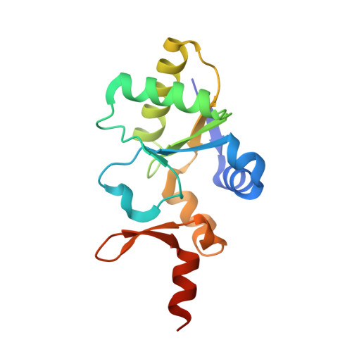
Structural characterization of the As/Sb reductase LmACR2 from Leishmania major.
Mukhopadhyay, R., Bisacchi, D., Zhou, Y., Armirotti, A., Bordo, D.(2009) J Mol Biol 386: 1229-1239
- PubMed: 18687336
- DOI: https://doi.org/10.1016/j.jmb.2008.07.056
- Primary Citation of Related Structures:
2J6P - PubMed Abstract:
The arsenate/antimonate reductase LmACR2 has been recently identified in the genome of Leishmania major. Besides displaying phosphatase activity in vitro, this enzyme is able to reduce both As(V) and Sb(V) to their respective trivalent forms and is involved in the activation of Pentostan, a drug containing Sb(V) used in the treatment of leishmaniasis. LmACR2 displays sequence and functional similarity with the arsenate reductase ScACR2 from Saccharomyces cerevisiae, and both proteins are homologous to the catalytic domain of Cdc25 phosphatases, which, in turn, belong to the rhodanese/Cdc25 phosphatase superfamily. In this work, the three-dimensional structure of LmACR2 has been determined with crystallographic methods and refined at 2.15 A resolution. The protein structure maintains the overall rhodanese fold, but substantial modifications are observed in secondary structure position and length. However, the conformation of the active-site loop and the position of the catalytic residue Cys75 are unchanged with respect to the Cdc25 phosphatases. From an evolutionary viewpoint, LmACR2 and the related arsenate reductases form, together with the known Cdc25 phosphatases, a well-defined subfamily of the rhodanese/Cdc25 phosphatase superfamily, characterized by a 7-amino-acid-long active-site loop that is able to selectively bind substrates containing phosphorous, arsenic, or antinomy. The evolutionary tree obtained for these proteins shows that, besides the active-site motif CE[F/Y]SXXR that characterizes Cdc25 phosphatase, the novel CALSQ[Q/V]R motif is also conserved in sequences from fungi and plants. Similar to Cdc25 phosphatase, these proteins are likely involved in cell cycle control. The active-site composition of LmACR2 (CAQSLVR) does not belong to either group, but gives to the enzyme a bifunctional activity of both phosphatase and As/Sb reductase. The subtle dependence of substrate specificity on the amino acid composition of the active-site loop displays the versatility of the ubiquitous rhodanese domain.
Organizational Affiliation:
Department of Biochemistry and Molecular Biology, School of Medicine, Wayne State University, 540 East Canfield Avenue, Detroit, MI 48201, USA.