2I4P

Crystal structure of the complex between PPARgamma and the partial agonist LT127 (ureidofibrate derivative). Structure obtained from crystals of the apo-form soaked for 30 days.


Experimental Data Snapshot

  • Method: X-RAY DIFFRACTION
  • Resolution: 2.10 Å
  • R-Value Free: 0.295 
  • R-Value Work: 0.272 
  • R-Value Observed: 0.273 

wwPDB Validation   3D Report Full Report


Ligand Structure Quality Assessment 


This is version 1.4 of the entry. See complete history


Literature

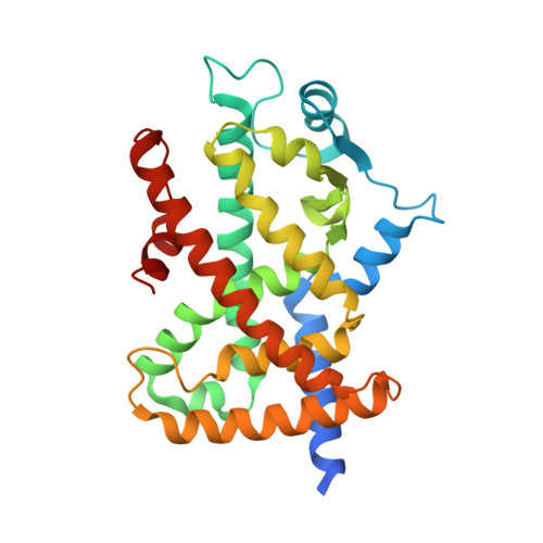
Insights into the mechanism of partial agonism: crystal structures of the peroxisome proliferator-activated receptor gamma ligand-binding domain in the complex with two enantiomeric ligands.

Pochetti, G.Godio, C.Mitro, N.Caruso, D.Galmozzi, A.Scurati, S.Loiodice, F.Fracchiolla, G.Tortorella, P.Laghezza, A.Lavecchia, A.Novellino, E.Mazza, F.Crestani, M.

(2007) J Biol Chem 282: 17314-17324

  • DOI: https://doi.org/10.1074/jbc.M702316200
  • Primary Citation of Related Structures:  
    2I4J, 2I4P, 2I4Z

  • PubMed Abstract: 

    The peroxisome proliferator-activated receptors (PPARs) are transcriptional regulators of glucose and lipid metabolism. They are activated by natural ligands, such as fatty acids, and are also targets of synthetic antidiabetic and hypolipidemic drugs. By using cell-based reporter assays, we studied the transactivation activity of two enantiomeric ureidofibrate-like derivatives. In particular, we show that the R-enantiomer, (R)-1, is a full agonist of PPARgamma, whereas the S-enantiomer, (S)-1, is a less potent partial agonist. Most importantly, we report the x-ray crystal structures of the PPARgamma ligand binding domain complexed with the R- and the S-enantiomer, respectively. The analysis of the two crystal structures shows that the different degree of stabilization of the helix 12 induced by the ligand determines its behavior as full or partial agonist. Another crystal structure of the PPARgamma.(S)-1 complex, only differing in the soaking time of the ligand, is also presented. The comparison of the two structures of the complexes with the partial agonist reveals significant differences and is suggestive of the possible coexistence in solution of transcriptionally active and inactive forms of helix 12 in the presence of a partial agonist. Mutation analysis confirms the importance of Leu(465), Leu(469), and Ile(472) in the activation by (R)-1 and underscores the key role of Gln(286) in the PPARgamma activity.


  • Organizational Affiliation

    Istituto di Cristallografia, Consiglio Nazionale delle Ricerche, Montelibretti, 00016 Monterotondo Stazione, Roma, Italia. giorgio.pochetti@ic.cnr.it


Macromolecules
Find similar proteins by:  (by identity cutoff)  |  3D Structure
Entity ID: 1
MoleculeChains Sequence LengthOrganismDetailsImage
Peroxisome proliferator-activated receptor gamma
A, B
286Homo sapiensMutation(s): 0 
Gene Names: PPARG
UniProt & NIH Common Fund Data Resources
Find proteins for P37231 (Homo sapiens)
Explore P37231 
Go to UniProtKB:  P37231
PHAROS:  P37231
GTEx:  ENSG00000132170 
Entity Groups  
Sequence Clusters30% Identity50% Identity70% Identity90% Identity95% Identity100% Identity
UniProt GroupP37231
Sequence Annotations
Expand
  • Reference Sequence
Small Molecules
Ligands 1 Unique
IDChains Name / Formula / InChI Key2D Diagram3D Interactions
DRH
Query on DRH

Download Ideal Coordinates CCD File 
C [auth A](2S)-2-(4-{2-[1,3-BENZOXAZOL-2-YL(HEPTYL)AMINO]ETHYL}PHENOXY)-2-METHYLBUTANOIC ACID
C27 H36 N2 O4
QPKIEBNVIOELIR-MHZLTWQESA-N
Binding Affinity Annotations 
IDSourceBinding Affinity
DRH BindingDB:  2I4P Ki: 971 (nM) from 1 assay(s)
Kd: min: 1978, max: 2000 (nM) from 2 assay(s)
EC50: min: 590, max: 593 (nM) from 2 assay(s)
PDBBind:  2I4P Kd: 1978 (nM) from 1 assay(s)
Experimental Data & Validation

Experimental Data

  • Method: X-RAY DIFFRACTION
  • Resolution: 2.10 Å
  • R-Value Free: 0.295 
  • R-Value Work: 0.272 
  • R-Value Observed: 0.273 
  • Space Group: C 1 2 1
Unit Cell:
Length ( Å )Angle ( ˚ )
a = 93.54α = 90
b = 60.91β = 103.08
c = 118.35γ = 90
Software Package:
Software NamePurpose
DENZOdata reduction
SCALEPACKdata scaling
CNSrefinement
PDB_EXTRACTdata extraction
MAR345data collection
CNSphasing

Structure Validation

View Full Validation Report


Ligand Structure Quality Assessment 


Entry History 

Deposition Data

Revision History  (Full details and data files)

  • Version 1.0: 2007-04-17
    Type: Initial release
  • Version 1.1: 2008-05-01
    Changes: Version format compliance
  • Version 1.2: 2011-07-13
    Changes: Version format compliance
  • Version 1.3: 2017-10-18
    Changes: Advisory, Refinement description
  • Version 1.4: 2024-02-21
    Changes: Advisory, Data collection, Database references, Derived calculations