


Structure of the C-terminal domain of DipZ from Mycobacterium tuberculosis
Goldstone, D., Baker, E.N., Metcalf, P.To be published.
Experimental Data Snapshot
wwPDB Validation 3D Report Full Report

Entity ID: 1 | |||||
|---|---|---|---|---|---|
| Molecule | Chains | Sequence Length | Organism | Details | Image |
| Protein dipZ | 352 | Mycobacterium tuberculosis H37Rv | Mutation(s): 0 Gene Names: dipZ | 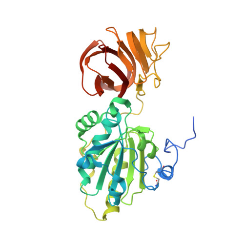 | |
UniProt | |||||
Find proteins for P9WG63 (Mycobacterium tuberculosis (strain ATCC 25618 / H37Rv)) Explore P9WG63 Go to UniProtKB: P9WG63 | |||||
Entity Groups | |||||
| Sequence Clusters | 30% Identity50% Identity70% Identity90% Identity95% Identity100% Identity | ||||
| UniProt Group | P9WG63 | ||||
Sequence AnnotationsExpand | |||||
| |||||
| Length ( Å ) | Angle ( ˚ ) |
|---|---|
| a = 110.07 | α = 90 |
| b = 117.915 | β = 90 |
| c = 123.03 | γ = 90 |
| Software Name | Purpose |
|---|---|
| DENZO | data reduction |
| SCALEPACK | data scaling |
| SOLVE | phasing |
| RESOLVE | phasing |
| CNS | refinement |
| PDB_EXTRACT | data extraction |
| Blu-Ice | data collection |
RCSB PDB (citation) is hosted by
RCSB PDB is a member of the

