Three-dimensional structure of the human immunodeficiency virus type 1 matrix protein.
Massiah, M.A., Starich, M.R., Paschall, C., Summers, M.F., Christensen, A.M., Sundquist, W.I.(1994) J Mol Biol 244: 198-223
- PubMed: 7966331
- DOI: https://doi.org/10.1006/jmbi.1994.1719
- Primary Citation of Related Structures:
2HMX - PubMed Abstract:
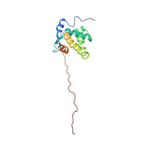
The HIV-1 matrix protein forms an icosahedral shell associated with the inner membrane of the mature virus. Genetic analyses have indicated that the protein performs important functions throughout the viral life-cycle, including anchoring the transmembrane envelope protein on the surface of the virus, assisting in viral penetration, transporting the proviral integration complex across the nuclear envelope, and localizing the assembling virion to the cell membrane. We now report the three-dimensional structure of recombinant HIV-1 matrix protein, determined at high resolution by nuclear magnetic resonance (NMR) methods. The HIV-1 matrix protein is the first retroviral matrix protein to be characterized structurally and only the fourth HIV-1 protein of known structure. NMR signal assignments required recently developed triple-resonance (1H, 13C, 15N) NMR methodologies because signals for 91% of 132 assigned H alpha protons and 74% of the 129 assignable backbone amide protons resonate within chemical shift ranges of 0.8 p.p.m. and 1 p.p.m., respectively. A total of 636 nuclear Overhauser effect-derived distance restraints were employed for distance geometry-based structure calculations, affording an average of 13.0 NMR-derived distance restraints per residue for the experimentally constrained amino acids. An ensemble of 25 refined distance geometry structures with penalties (sum of the squares of the distance violations) of 0.32 A2 or less and individual distance violations under 0.06 A was generated; best-fit superposition of ordered backbone heavy atoms relative to mean atom positions afforded root-mean-square deviations of 0.50 (+/- 0.08) A. The folded HIV-1 matrix protein structure is composed of five alpha-helices, a short 3(10) helical stretch, and a three-strand mixed beta-sheet. Helices I to III and the 3(10) helix pack about a central helix (IV) to form a compact globular domain that is capped by the beta-sheet. The C-terminal helix (helix V) projects away from the beta-sheet to expose carboxyl-terminal residues essential for early steps in the HIV-1 infectious cycle. Basic residues implicated in membrane binding and nuclear localization functions cluster about an extruded cationic loop that connects beta-strands 1 and 2. The structure suggests that both membrane binding and nuclear localization may be mediated by complex tertiary structures rather than simple linear determinants.
Organizational Affiliation:
Howard Hughes Medical Institute, Baltimore, MD.