2H44

Crystal structure of PDE5A1 in complex with icarisid II


Experimental Data Snapshot

  • Method: X-RAY DIFFRACTION
  • Resolution: 1.80 Å
  • R-Value Free: 0.233 
  • R-Value Work: 0.206 

wwPDB Validation   3D Report Full Report


This is version 1.3 of the entry. See complete history


Literature

Multiple Conformations of Phosphodiesterase-5: Implications for enzyme function and drug development

Wang, H.Liu, Y.Huai, Q.Cai, J.Zoraghi, R.Francis, S.H.Corbin, J.D.Robinson, H.Xin, Z.Lin, G.Ke, H.

(2006) J Biol Chem 281: 21469-21479

  • DOI: https://doi.org/10.1074/jbc.M512527200
  • Primary Citation of Related Structures:  
    2H40, 2H42, 2H44

  • PubMed Abstract: 

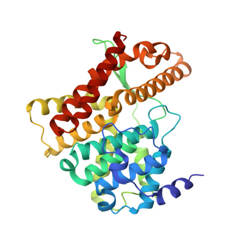
    Phosphodiesterase-5 (PDE5) is the target for sildenafil, vardenafil, and tadalafil, which are drugs for treatment of erectile dysfunction and pulmonary hypertension. We report here the crystal structures of a fully active catalytic domain of unliganded PDE5A1 and its complexes with sildenafil or icarisid II. These structures together with the PDE5A1-isobutyl-1-methylxanthine complex show that the H-loop (residues 660-683) at the active site of PDE5A1 has four different conformations and migrates 7-35A upon inhibitor binding. In addition, the conformation of sildenafil reported herein differs significantly from those in the previous structures of chimerically hybridized or almost inactive PDE5. Mutagenesis and kinetic analyses confirm that the H-loop is particularly important for substrate recognition and that invariant Gly(659), which immediately precedes the H-loop, is critical for optimal substrate affinity and catalytic activity.


  • Organizational Affiliation

    Department of Biochemistry and Biophysics and Lineberger Comprehensive Cancer Center, University of North Carolina, Chapel Hill, North Carolina 27599-7260.


Macromolecules
Find similar proteins by:  (by identity cutoff)  |  3D Structure
Entity ID: 1
MoleculeChains Sequence LengthOrganismDetailsImage
cGMP-specific 3',5'-cyclic phosphodiesterase326Homo sapiensMutation(s): 0 
Gene Names: PDE5APDE5
EC: 3.1.4.35
UniProt & NIH Common Fund Data Resources
Find proteins for O76074 (Homo sapiens)
Explore O76074 
Go to UniProtKB:  O76074
PHAROS:  O76074
GTEx:  ENSG00000138735 
Entity Groups  
Sequence Clusters30% Identity50% Identity70% Identity90% Identity95% Identity100% Identity
UniProt GroupO76074
Sequence Annotations
Expand
  • Reference Sequence
Small Molecules
Ligands 3 Unique
IDChains Name / Formula / InChI Key2D Diagram3D Interactions
7CA
Query on 7CA

Download Ideal Coordinates CCD File 
D [auth A]5,7-DIHYDROXY-2-(4-METHOXYPHENYL)-8-(3-METHYLBUTYL)-4-OXO-4H-CHROMEN-3-YL 6-DEOXY-ALPHA-L-MANNOPYRANOSIDE
C27 H32 O10
PFVZUXQCELCLBL-LVKFHIPRSA-N
ZN
Query on ZN

Download Ideal Coordinates CCD File 
B [auth A]ZINC ION
Zn
PTFCDOFLOPIGGS-UHFFFAOYSA-N
MG
Query on MG

Download Ideal Coordinates CCD File 
C [auth A]MAGNESIUM ION
Mg
JLVVSXFLKOJNIY-UHFFFAOYSA-N
Binding Affinity Annotations 
IDSourceBinding Affinity
7CA BindingDB:  2H44 IC50: min: 1700, max: 5.50e+4 (nM) from 3 assay(s)
PDBBind:  2H44 IC50: 1700 (nM) from 1 assay(s)
Binding MOAD:  2H44 IC50: 1700 (nM) from 1 assay(s)
Experimental Data & Validation

Experimental Data

  • Method: X-RAY DIFFRACTION
  • Resolution: 1.80 Å
  • R-Value Free: 0.233 
  • R-Value Work: 0.206 
  • Space Group: P 61 2 2
Unit Cell:
Length ( Å )Angle ( ˚ )
a = 110.746α = 90
b = 110.746β = 90
c = 106.159γ = 120
Software Package:
Software NamePurpose
ADSCdata collection
HKL-2000data reduction
AMoREphasing
CNSrefinement
HKL-2000data scaling

Structure Validation

View Full Validation Report


Entry History 

Deposition Data

Revision History  (Full details and data files)

  • Version 1.0: 2006-06-06
    Type: Initial release
  • Version 1.1: 2008-05-01
    Changes: Version format compliance
  • Version 1.2: 2011-07-13
    Changes: Version format compliance
  • Version 1.3: 2023-08-30
    Changes: Data collection, Database references, Derived calculations, Refinement description