2GLI

FIVE-FINGER GLI/DNA COMPLEX


Experimental Data Snapshot

  • Method: X-RAY DIFFRACTION
  • Resolution: 2.60 Å
  • R-Value Observed: 0.228 

wwPDB Validation   3D Report Full Report


This is version 1.3 of the entry. See complete history


Literature

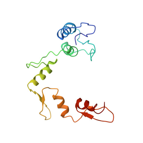
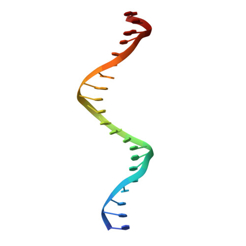
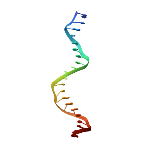
Crystal structure of a five-finger GLI-DNA complex: new perspectives on zinc fingers.

Pavletich, N.P.Pabo, C.O.

(1993) Science 261: 1701-1707

  • DOI: https://doi.org/10.1126/science.8378770
  • Primary Citation of Related Structures:  
    2GLI

  • PubMed Abstract: 

    Zinc finger proteins, of the type first discovered in transcription factor IIIA (TFIIIA), are one of the largest and most important families of DNA-binding proteins. The crystal structure of a complex containing the five Zn fingers from the human GLI oncogene and a high-affinity DNA binding site has been determined at 2.6 A resolution. Finger one does not contact the DNA. Fingers two through five bind in the major groove and wrap around the DNA, but lack the simple, strictly periodic arrangement observed in the Zif268 complex. Fingers four and five of GLI make extensive base contacts in a conserved nine base-pair region, and this section of the DNA has a conformation intermediate between B-DNA and A-DNA. Analyzing the GLI complex and comparing it with Zif268 offers new perspectives on Zn finger-DNA recognition.


  • Organizational Affiliation

    Howard Hughes Medical Institute, Massachusetts Institute of Technology, Cambridge 02139.


Macromolecules

Find similar proteins by:  (by identity cutoff)  |  3D Structure
Entity ID: 3
MoleculeChains Sequence LengthOrganismDetailsImage
PROTEIN (FIVE-FINGER GLI)C [auth A]155Homo sapiensMutation(s): 0 
Gene Names: GLI ONCOGENE
UniProt & NIH Common Fund Data Resources
Find proteins for P08151 (Homo sapiens)
Explore P08151 
Go to UniProtKB:  P08151
PHAROS:  P08151
GTEx:  ENSG00000111087 
Entity Groups  
Sequence Clusters30% Identity50% Identity70% Identity90% Identity95% Identity100% Identity
UniProt GroupP08151
Sequence Annotations
Expand
  • Reference Sequence

Find similar nucleic acids by:  Sequence   |   3D Structure  

Entity ID: 1
MoleculeChains LengthOrganismImage
DNA (5'-D(*TP*TP*TP*CP*GP*TP*CP*TP*TP*GP*GP*GP*TP*GP*GP*TP*CP*CP*AP*CP*G)-3')A [auth C]21N/A
Sequence Annotations
Expand
  • Reference Sequence

Find similar nucleic acids by:  Sequence   |   3D Structure  

Entity ID: 2
MoleculeChains LengthOrganismImage
DNA (5'-D(*AP*CP*GP*TP*GP*GP*AP*CP*CP*AP*CP*CP*CP*AP*AP*GP*AP*CP*GP*AP*A)-3')B [auth D]21N/A
Sequence Annotations
Expand
  • Reference Sequence
Experimental Data & Validation

Experimental Data

  • Method: X-RAY DIFFRACTION
  • Resolution: 2.60 Å
  • R-Value Observed: 0.228 
  • Space Group: P 21 21 21
Unit Cell:
Length ( Å )Angle ( ˚ )
a = 149α = 90
b = 50.2β = 90
c = 45.4γ = 90
Software Package:
Software NamePurpose
TNTrefinement

Structure Validation

View Full Validation Report


Entry History 

Deposition Data

Revision History  (Full details and data files)

  • Version 1.0: 1993-11-09
    Type: Initial release
  • Version 1.1: 2008-05-22
    Changes: Version format compliance
  • Version 1.2: 2011-07-13
    Changes: Version format compliance
  • Version 1.3: 2024-02-14
    Changes: Data collection, Database references, Derived calculations