2GEJ

Crystal Structure of phosphatidylinositol mannosyltransferase (PimA) from Mycobacterium smegmatis in complex with GDP-Man


Experimental Data Snapshot

  • Method: X-RAY DIFFRACTION
  • Resolution: 2.60 Å
  • R-Value Free: 0.245 
  • R-Value Work: 0.192 
  • R-Value Observed: 0.196 

wwPDB Validation   3D Report Full Report


This is version 1.3 of the entry. See complete history


Literature

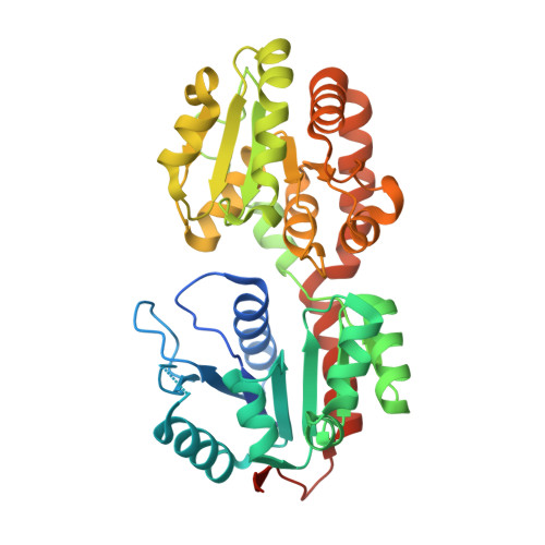
Molecular recognition and interfacial catalysis by the essential phosphatidylinositol mannosyltransferase PimA from mycobacteria.

Guerin, M.E.Kordulakova, J.Schaeffer, F.Svetlikova, Z.Buschiazzo, A.Giganti, D.Gicquel, B.Mikusova, K.Jackson, M.Alzari, P.M.

(2007) J Biol Chem 282: 20705-20714

  • DOI: https://doi.org/10.1074/jbc.M702087200
  • Primary Citation of Related Structures:  
    2GEJ, 2GEK

  • PubMed Abstract: 

    Mycobacterial phosphatidylinositol mannosides (PIMs) and metabolically derived cell wall lipoglycans play important roles in host-pathogen interactions, but their biosynthetic pathways are poorly understood. Here we focus on Mycobacterium smegmatis PimA, an essential enzyme responsible for the initial mannosylation of phosphatidylinositol. The structure of PimA in complex with GDP-mannose shows the two-domain organization and the catalytic machinery typical of GT-B glycosyltransferases. PimA is an amphitrophic enzyme that binds mono-disperse phosphatidylinositol, but its transferase activity is stimulated by high concentrations of non-substrate anionic surfactants, indicating that the early stages of PIM biosynthesis involve lipid-water interfacial catalysis. Based on structural, calorimetric, and mutagenesis studies, we propose a model wherein PimA attaches to the membrane through its N-terminal domain, and this association leads to enzyme activation. Our results reveal a novel mode of phosphatidylinositol recognition and provide a template for the development of potential antimycobacterial compounds.


  • Organizational Affiliation

    Unité de Biochimie Structurale (CNRS URA 2185), Institut Pasteur, 25 rue du Docteur Roux, 75724 Paris Cedex 15, France.


Macromolecules
Find similar proteins by:  (by identity cutoff)  |  3D Structure
Entity ID: 1
MoleculeChains Sequence LengthOrganismDetailsImage
PHOSPHATIDYLINOSITOL MANNOSYLTRANSFERASE (PimA)406Mycolicibacterium smegmatis MC2 155Mutation(s): 0 
Gene Names: PIMA
UniProt
Find proteins for A0QWG6 (Mycolicibacterium smegmatis (strain ATCC 700084 / mc(2)155))
Explore A0QWG6 
Go to UniProtKB:  A0QWG6
Entity Groups  
Sequence Clusters30% Identity50% Identity70% Identity90% Identity95% Identity100% Identity
UniProt GroupA0QWG6
Sequence Annotations
Expand
  • Reference Sequence
Small Molecules
Ligands 1 Unique
IDChains Name / Formula / InChI Key2D Diagram3D Interactions
GDD
Query on GDD

Download Ideal Coordinates CCD File 
B [auth A]GUANOSINE-5'-DIPHOSPHATE-ALPHA-D-MANNOSE
C16 H25 N5 O16 P2
MVMSCBBUIHUTGJ-GDJBGNAASA-N
Binding Affinity Annotations 
IDSourceBinding Affinity
GDD PDBBind:  2GEJ Kd: 230 (nM) from 1 assay(s)
Experimental Data & Validation

Experimental Data

  • Method: X-RAY DIFFRACTION
  • Resolution: 2.60 Å
  • R-Value Free: 0.245 
  • R-Value Work: 0.192 
  • R-Value Observed: 0.196 
  • Space Group: P 21 21 21
Unit Cell:
Length ( Å )Angle ( ˚ )
a = 37.071α = 90
b = 72.747β = 90
c = 137.166γ = 90
Software Package:
Software NamePurpose
SCALAdata scaling
AMoREphasing
REFMACrefinement
PDB_EXTRACTdata extraction
MOSFLMdata reduction
CCP4data scaling

Structure Validation

View Full Validation Report


Entry History 

Deposition Data

Revision History  (Full details and data files)

  • Version 1.0: 2007-04-03
    Type: Initial release
  • Version 1.1: 2008-05-01
    Changes: Version format compliance
  • Version 1.2: 2011-07-13
    Changes: Source and taxonomy, Version format compliance
  • Version 1.3: 2024-02-14
    Changes: Data collection, Database references, Derived calculations