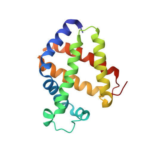
Cyanide binding and heme cavity conformational transitions in Drosophila melanogaster hexacoordinate hemoglobin.
de Sanctis, D., Ascenzi, P., Bocedi, A., Dewilde, S., Burmester, T., Hankeln, T., Moens, L., Bolognesi, M.(2006) Biochemistry 45: 10054-10061
- PubMed: 16906763
- DOI: https://doi.org/10.1021/bi060462a
- Primary Citation of Related Structures:
2G3H - PubMed Abstract:
The reason for the presence of hemoglobin-like molecules in insects, such as Drosophila melanogaster, that live in fully aerobic environments has yet to be determined. Heme endogenous hexacoordination (where HisE7 and HisF8 axial ligands to the heme Fe atom are both provided by the protein) is a recently discovered mechanism proposed to modulate O(2) affinity in hemoglobins from different species. Previous results have shown that D. melanogaster hemoglobin 1 (product of the glob1 gene) displays heme endogenous hexacoordination in both the ferrous and ferric states. Here we present kinetic data characterizing the exogenous cyanide ligand binding process, and the three-dimensional structure (at 1.4 A resolution) of the ensuing cyano-met D. melanogaster hemoglobin. Comparison with the crystal structure of the endogenously hexacoordinated D. melanogaster hemoglobin shows that the transition to the cyano-met form is supported by conformational readjustment in the CD-D-E region of the protein, which removes HisE7 from the heme. The structural and functional features of D. melanogaster hemoglobin are examined in light of previous results achieved for human and mouse neuroglobins and for human cytoglobin, which display heme endogenous hexacoordination. The study shows that, despite the rather constant value for cyanide association rate constants for the ferric hemoproteins, different distal site conformational readjustments and/or heme sliding mechanisms are displayed by the known hexacoordinate hemoglobins as a result of exogenous ligand binding.
Organizational Affiliation:
Instituto de Tecnologia Quimica e Biologica, Universidade Nova de Lisboa, P.O. Box 127, 2781-901 Oeiras, Portugal.