2FUS

MUTATIONS OF FUMARASE THAT DISTINGUISH BETWEEN THE ACTIVE SITE AND A NEARBY DICARBOXYLIC ACID BINDING SITE


Experimental Data Snapshot

  • Method: X-RAY DIFFRACTION
  • Resolution: 2.20 Å
  • R-Value Free: 0.229 
  • R-Value Work: 0.178 
  • R-Value Observed: 0.178 

wwPDB Validation   3D Report Full Report


This is version 1.4 of the entry. See complete history


Literature

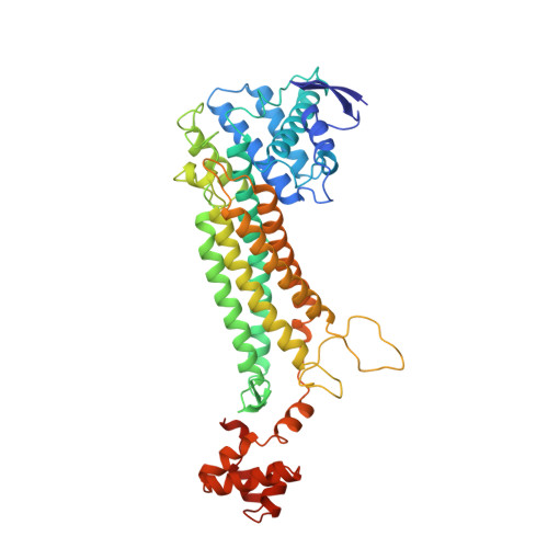
Mutations of fumarase that distinguish between the active site and a nearby dicarboxylic acid binding site.

Weaver, T.Lees, M.Banaszak, L.

(1997) Protein Sci 6: 834-842

  • DOI: https://doi.org/10.1002/pro.5560060410
  • Primary Citation of Related Structures:  
    1FUR, 2FUS

  • PubMed Abstract: 

    Two mutant forms of fumarase C from E. coli have been made using PCR and recombinant DNA. The recombinant form of the protein included a histidine arm on the C-terminal facilitating purification. Based on earlier studies, two different carboxylic acid binding sites, labeled A- and B-, were observed in crystal structures of the wild type and inhibited forms of the enzyme. A histidine at each of the sites was mutated to an asparagine. H188N at the A-site resulted in a large decrease in specific activity, while the H129N mutation at the B-site had essentially no effect. From the results, we conclude that the A-site is indeed the active site, and a dual role for H188 as a potential catalytic base is proposed. Crystal structures of the two mutant proteins produced some unexpected results. Both mutations reduced the affinity for the carboxylic acids at their respective sites. The H129N mutant should be particularly useful in future kinetic studies because it sterically blocks the B-site with the carboxyamide of asparagine assuming the position of the ligand's carboxylate. In the H188N mutation at the active site, the new asparagine side chain still interacts with an active site water that appears to have moved slightly as a result of the mutation.


  • Organizational Affiliation

    Department of Biochemistry, University of Minnesota, Minneapolis 55455, USA.


Macromolecules
Find similar proteins by:  (by identity cutoff)  |  3D Structure
Entity ID: 1
MoleculeChains Sequence LengthOrganismDetailsImage
FUMARASE C
A, B
467Escherichia coliMutation(s): 1 
Gene Names: FUMC
EC: 4.2.1.2
UniProt
Find proteins for P05042 (Escherichia coli (strain K12))
Explore P05042 
Go to UniProtKB:  P05042
Entity Groups  
Sequence Clusters30% Identity50% Identity70% Identity90% Identity95% Identity100% Identity
UniProt GroupP05042
Sequence Annotations
Expand
  • Reference Sequence
Experimental Data & Validation

Experimental Data

  • Method: X-RAY DIFFRACTION
  • Resolution: 2.20 Å
  • R-Value Free: 0.229 
  • R-Value Work: 0.178 
  • R-Value Observed: 0.178 
  • Space Group: C 2 2 21
Unit Cell:
Length ( Å )Angle ( ˚ )
a = 104.16α = 90
b = 220.05β = 90
c = 86.65γ = 90
Software Package:
Software NamePurpose
X-PLORmodel building
X-PLORrefinement
XENGENdata reduction
XENGENdata scaling
X-PLORphasing

Structure Validation

View Full Validation Report


Entry History 

Deposition Data

Revision History  (Full details and data files)

  • Version 1.0: 1997-07-23
    Type: Initial release
  • Version 1.1: 2008-03-03
    Changes: Version format compliance
  • Version 1.2: 2011-07-13
    Changes: Derived calculations, Version format compliance
  • Version 1.3: 2021-11-03
    Changes: Database references, Derived calculations, Other
  • Version 1.4: 2023-08-09
    Changes: Refinement description