2E67

Crystal structure of the hypothetical protein TTHB029 from Thermus thermophilus HB8


Experimental Data Snapshot

  • Method: X-RAY DIFFRACTION
  • Resolution: 2.90 Å
  • R-Value Free: 0.259 
  • R-Value Work: 0.219 
  • R-Value Observed: 0.221 

wwPDB Validation   3D Report Full Report


This is version 1.3 of the entry. See complete history


Literature

Crystal structure of the YdjC-family protein TTHB029 from Thermus thermophilus HB8: structural relationship with peptidoglycan N-acetylglucosamine deacetylase.

Imagawa, T.Iino, H.Kanagawa, M.Ebihara, A.Kuramitsu, S.Tsuge, H.

(2008) Biochem Biophys Res Commun 367: 535-541

  • DOI: https://doi.org/10.1016/j.bbrc.2007.12.144
  • Primary Citation of Related Structures:  
    2E67

  • PubMed Abstract: 

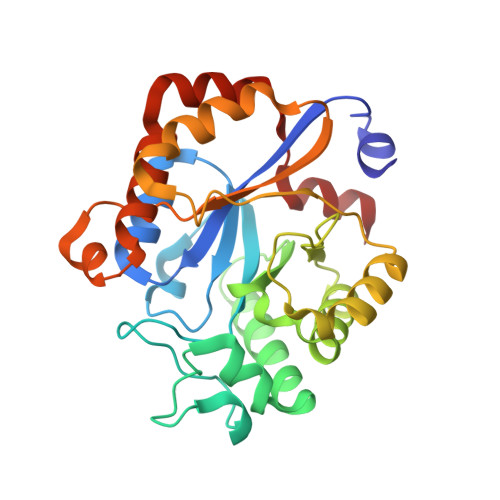
    The YdjC-family protein is widely distributed, from human to bacteria, but so far no three-dimensional structure and functional analysis of this family of proteins has been reported. We determined the three-dimensional structure of the YdjC homolog TTHB029 at a resolution of 2.9A. The overall structure of the monomer consists of (betaalpha)-barrel fold forming a homodimer. Asp21, His60, and His127 residues coordinate to Mg(2+) as a possible active site. TTHB029 shows structural similarity to the peptidoglycan N-acetylglucosamine deacetylase from Streptococcus pneumoniae (SpPgdA). The active site groove of SpPgdA includes the Zn(2+) coordinated to Asp276, His326, and His330. Despite the low sequence identity, metal-binding residues of Asp-His-His were conserved among the two enzymes. There were definitive differences, however, in that one of the histidines of the metal-binding site was substituted for the other histidine located on the other loop. Moreover, these important metal-binding residues and the residues of the presumed active site are fully conserved in YdjC-family protein.


  • Organizational Affiliation

    Institute for Health Sciences, Tokushima Bunri University, 180 Nishihama, Yamashiro, Tokushima 770-8514, Japan.


Macromolecules
Find similar proteins by:  (by identity cutoff)  |  3D Structure
Entity ID: 1
MoleculeChains Sequence LengthOrganismDetailsImage
Hypothetical protein TTHB029
A, B, C, D, E
A, B, C, D, E, F
264Thermus thermophilus HB8Mutation(s): 0 
UniProt
Find proteins for Q53WD3 (Thermus thermophilus (strain ATCC 27634 / DSM 579 / HB8))
Explore Q53WD3 
Go to UniProtKB:  Q53WD3
Entity Groups  
Sequence Clusters30% Identity50% Identity70% Identity90% Identity95% Identity100% Identity
UniProt GroupQ53WD3
Sequence Annotations
Expand
  • Reference Sequence
Experimental Data & Validation

Experimental Data

  • Method: X-RAY DIFFRACTION
  • Resolution: 2.90 Å
  • R-Value Free: 0.259 
  • R-Value Work: 0.219 
  • R-Value Observed: 0.221 
  • Space Group: H 3 2
Unit Cell:
Length ( Å )Angle ( ˚ )
a = 202.006α = 90
b = 202.006β = 90
c = 345.294γ = 120
Software Package:
Software NamePurpose
REFMACrefinement
HKL-2000data collection
HKL-2000data reduction
HKL-2000data scaling
SOLVEphasing

Structure Validation

View Full Validation Report


Entry History 

Deposition Data

Revision History  (Full details and data files)

  • Version 1.0: 2007-06-26
    Type: Initial release
  • Version 1.1: 2007-10-04
    Changes: Version format compliance
  • Version 1.2: 2011-07-13
    Changes: Source and taxonomy, Version format compliance
  • Version 1.3: 2024-03-13
    Changes: Data collection, Database references, Derived calculations