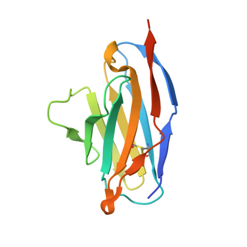
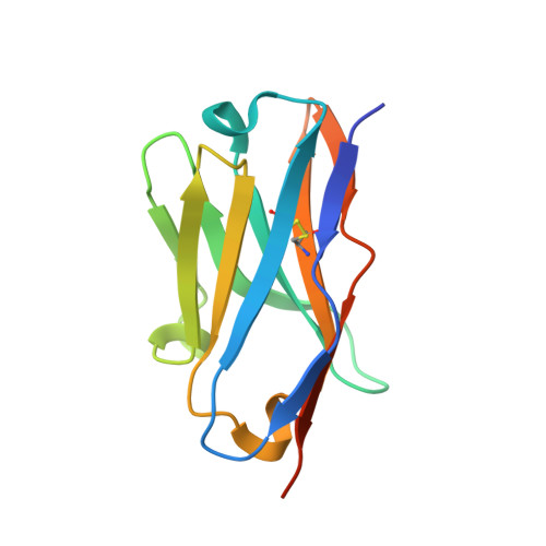
2E27

Crystal structure of Fv fragment of anti-ciguatoxin antibody complexed with ABC-ring of ciguatoxin


Experimental Data Snapshot

  • Method: X-RAY DIFFRACTION
  • Resolution: 1.70 Å
  • R-Value Free: 0.210 
  • R-Value Work: 0.198 
  • R-Value Observed: 0.198 

wwPDB Validation   3D Report Full Report


Ligand Structure Quality Assessment 


This is version 1.2 of the entry. See complete history


Literature

Critical contribution of aromatic rings to specific recognition of polyether rings. The case of ciguatoxin CTX3C-ABC and its specific antibody 1C49.

Tsumoto, K.Yokota, A.Tanaka, Y.Ui, M.Tsumuraya, T.Fujii, I.Kumagai, I.Nagumo, Y.Oguri, H.Inoue, M.Hirama, M.

(2008) J Biol Chem 283: 12259-12266

  • DOI: https://doi.org/10.1074/jbc.M710553200
  • Primary Citation of Related Structures:  
    2E27

  • PubMed Abstract: 

    To address how proteins recognize polyether toxin compounds, we focused on the interaction between the ABC ring compound of ciguatoxin 3C and its specific antibody, 1C49. Surface plasmon resonance analyses indicated that Escherichia coli-expressed variable domain fragments (Fv) of 1C49 had the high affinity constants and slow dissociation constants typical of antigen-antibody interactions. Linear van't Hoff analyses suggested that the interaction is enthalpy-driven. We resolved the crystal structure of 1C49 Fv bound to ABC ring compound of ciguatoxin 3C at a resolution of 1.7A. The binding pocket of the antibody had many aromatic rings and bound the antigen by shape complementarity typical of hapten-antibody interactions. Three hydrogen bonds and many van der Waals interactions were present. We mutated several residues of the antibody to Ala, and we used surface plasmon resonance to analyze the interactions between the mutated antibodies and the antigen. This analysis identified Tyr-91 and Trp-96 in the light chain as hot spots for the interaction, and other residues made incremental contributions by conferring enthalpic advantages and reducing the dissociation rate constant. Systematic mutation of Tyr-91 indicated that CH-pi and pi-pi interactions between the aromatic ring at this site and the antigen made substantial contributions to the association, and van der Waals interactions inhibited dissociation, suggesting that aromaticity and bulkiness are critical for the specific recognition of polyether compounds by proteins.


  • Organizational Affiliation

    Department of Medical Genome Sciences, Graduate School of Frontier Sciences, The University of Tokyo, Kashiwa 277-8562, Japan. tsumoto@k.u-tokyo.ac.jp


Macromolecules
Find similar proteins by:  (by identity cutoff)  |  3D Structure
Entity ID: 1
MoleculeChains Sequence LengthOrganismDetailsImage
anti-ciguatoxin antibody, light chainA [auth L]119Mus musculusMutation(s): 0 
Entity Groups  
Sequence Clusters30% Identity50% Identity70% Identity90% Identity95% Identity100% Identity
Sequence Annotations
Expand
  • Reference Sequence
Find similar proteins by:  (by identity cutoff)  |  3D Structure
Entity ID: 2
MoleculeChains Sequence LengthOrganismDetailsImage
anti-ciguatoxin antibody, heavy chainB [auth H]126Mus musculusMutation(s): 0 
Entity Groups  
Sequence Clusters30% Identity50% Identity70% Identity90% Identity95% Identity100% Identity
Sequence Annotations
Expand
  • Reference Sequence
Small Molecules
Ligands 3 Unique
IDChains Name / Formula / InChI Key2D Diagram3D Interactions
AB0
Query on AB0

Download Ideal Coordinates CCD File 
F [auth H]3,7:6,10:9,14-TRIANHYDRO-2,5,11,12,13-PENTADEOXY-4-O-(METHOXYMETHYL)-L-ARABINO-L-ALLO-TETRADEC-12-ENONIC ACID
C16 H24 O8
BUTGPEQQPCQQSZ-RQHZCWAZSA-N
GOL
Query on GOL

Download Ideal Coordinates CCD File 
D [auth L],
E [auth L],
H,
I [auth H],
J [auth H]
GLYCEROL
C3 H8 O3
PEDCQBHIVMGVHV-UHFFFAOYSA-N
DMS
Query on DMS

Download Ideal Coordinates CCD File 
C [auth L],
G [auth H]
DIMETHYL SULFOXIDE
C2 H6 O S
IAZDPXIOMUYVGZ-UHFFFAOYSA-N
Binding Affinity Annotations 
IDSourceBinding Affinity
AB0 Binding MOAD:  2E27 Ka: 2.22e+9 (M^-1) from 1 assay(s)
Experimental Data & Validation

Experimental Data

  • Method: X-RAY DIFFRACTION
  • Resolution: 1.70 Å
  • R-Value Free: 0.210 
  • R-Value Work: 0.198 
  • R-Value Observed: 0.198 
  • Space Group: I 21 21 21
Unit Cell:
Length ( Å )Angle ( ˚ )
a = 47.219α = 90
b = 97.502β = 90
c = 115.722γ = 90
Software Package:
Software NamePurpose
CNSrefinement
HKL-2000data collection
HKL-2000data reduction
SCALAdata scaling
CNSphasing

Structure Validation

View Full Validation Report


Ligand Structure Quality Assessment 


Entry History 

Deposition Data

Revision History  (Full details and data files)

  • Version 1.0: 2007-11-13
    Type: Initial release
  • Version 1.1: 2011-07-13
    Changes: Non-polymer description, Version format compliance
  • Version 1.2: 2023-10-25
    Changes: Data collection, Database references, Derived calculations, Refinement description